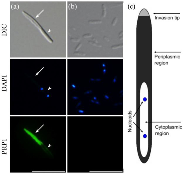
Figure 1.
Indirect immunofluorescence micrographs of H. obtusa with monoclonal antibodies mAb3D1B9C4 specific for the 63-kDa periplasmic protein PRP1. Left (a), IF; middle (b), RFs; right (c), a schematic representation of the IF of H. obtusa. Upper photos, differential interference contrast (DIC); middle photos, DAPI fluorescence; lower photos, AF488 immunofluorescence. Only a periplasmic region (arrow) except an invasion tip of the IF shows immunofluorescence. Because the cytoplasmic region of the IF is covered with a thin layer of the periplasm, a faint immunofluorescence layer appears around the cytoplasmic region. A cytoplasmic region (arrowhead) with two DAPI-positive nucleoids shows no immunofluorescence. H. obtusa cells on cover glasses and were permeabilized with 20 mM NaOH to allow antibodies entry inside the outer membrane. The RF shows no immunofluorescence. Scale bar, 10 µm.

