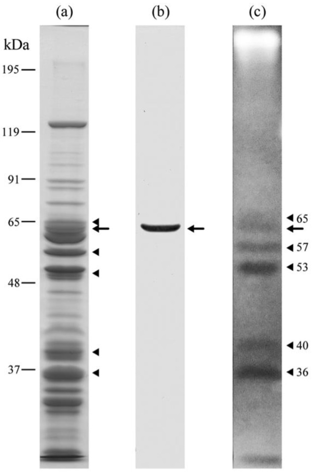
Figure 3.
SDS-DNA PAGE of IF cells of H. obtusa. Calf thymus DNA was used. (a) CBB stained gel; (b) immunoblot with mAb3D1B9C4 specific for PRP1; (c) EB stained gel. Arrows, PRP1. Arrowheads, other EB-staining negative bands showing positions of DNA-protein complexes (see Section 2).

