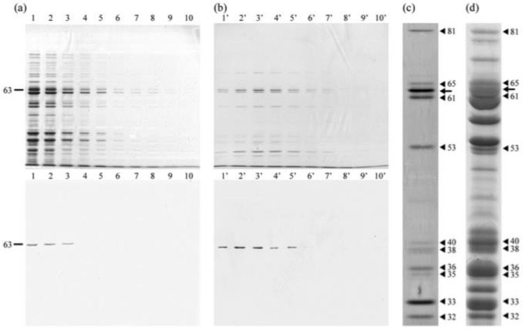
Figure 4.
SDS-PAGE and immunoblots of fractions of DNA affinity column chromatography. Calf thymus DNA was used. (a) Fractions 1–10 eluted with Na, K-PB; (b) fractions 1′–10′ eluted with Na, K-PB containing 0.2 M NaCl. Upper, silver-stained gel. Lower, immunoblot with mAb3D1B9C4. (c) High magnification of a fraction number 3′ using silver staining. Not only PRP1 but also several silver staining bands were also confirmed. (d) CBB stained gel of IF cells of H. obtusa. Arrow, PRP1. Arrowheads, other bands eluted. Bands in (c) match the bands in (d).

