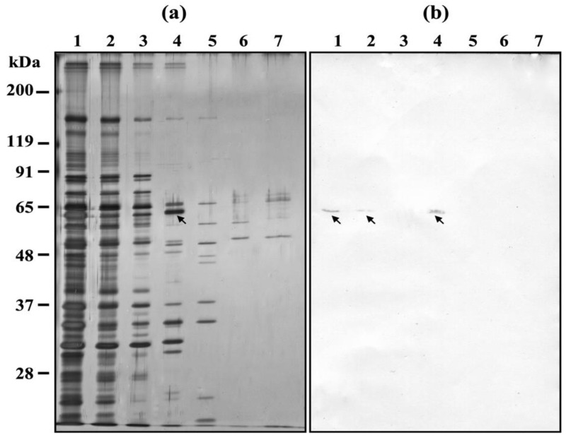
Figure 6.
DNA affinity chromatography of H. obtusa IFs. P. caudatum DNA was used. (a) Silver stained SDS-PAGE gel; (b) immunoblotting with the monoclonal antibody mAb3D1B9C4 specific for the 63-kDa periplasmic protein PRP1 of H. obtusa. Lane 1, supernatant of sonicated H. obtusa before applying to DNA affinity chromatography column; lane 2, first elute of supernatant of sonicated H. obtusa after applying to the column; lane 3, first elute by elution buffer containing 0.02 M NaCl; lane 4, first eluate by 0.1 M NaCl elution buffer; lane 5, first eluate by 0.2 M NaCl elution buffer; lane 6, first eluate by 0.3 M NaCl elution buffer; lane 7, first eluate by 2 M NaCl elution buffer; arrows, PRP1. Eluates from each column were collected in every 200 μL, and the first eluate from each column was used for SDS-PAGE and immunoblot with mAb. Note that not only PRP1 but also several silver stained bands were confirmed in lane 4 of (a).

