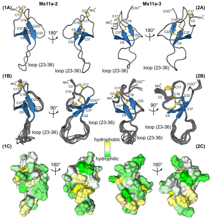
Figure 7.
Two-sided view on the Ms11a-2 (1) and Ms11a-3 (2) spatial structures. (A) Representative structures (with the fewest restraint violations) and (B) best 10 structures out of initial 100 superimposed over the backbone of β-sheet residues. Disulfide bonds are colored in yellow. (C) The contact surface of two molecules is colored according to the hydrophobicity, from yellow (hydrophobic) to green (hydrophilic) using the White and Wimley scale [30].

