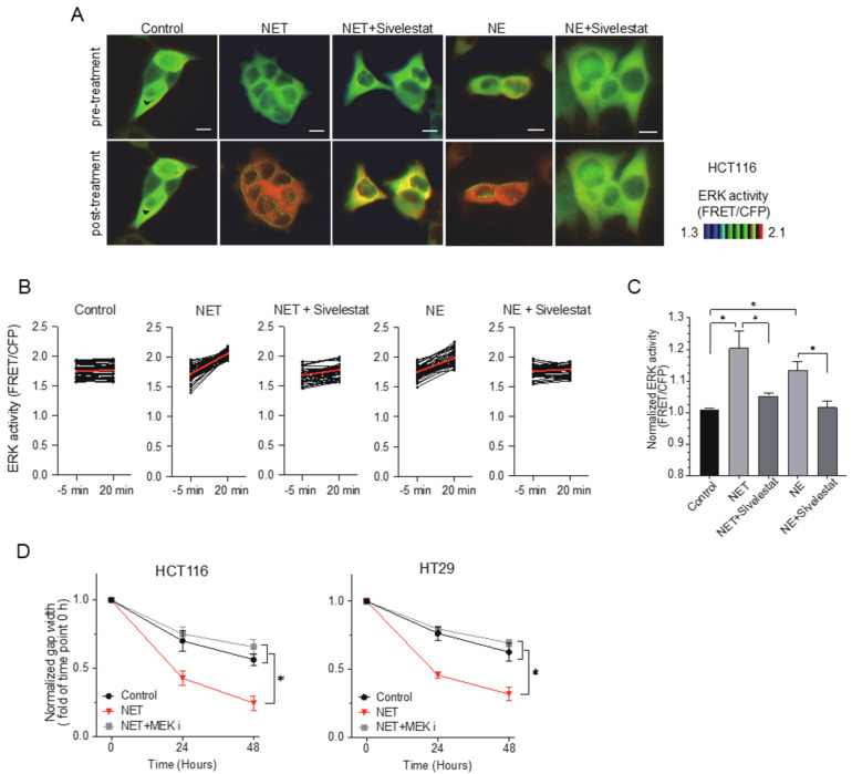
Figure 3.
Extracellular signal-regulated kinase (ERK) is an important regulator of activated cell migration induced by NETs. (A). Förster resonance energy transfer (FRET)-based live cell imaging using the biosensor of ERK in HCT116 cells. HCT116 cells were serum-starved for 6 hours. Cells incubated with NET-CM were treated with 10 µg/mL of NE or 100 µM of sivelestat. Representative FRET/CFP ratio images of pre-/post-treatment are shown in the intensity-modulated display mode. Scale bars, 10 μm. (B). ERK activity (FRET/CFP) of pre-/post-treatment in HCT116 cells. Each dot represents the ERK activity of each cell. A total of 54 cells were analyzed. Red bars indicate the mean values. (C) Increased rates in normalized ERK activity (FRET/CFP) of HCT116 cells before and after the addition of NET-CM. Results are presented as the means ± SEM of triplicate measurements. * p < 0.05 with Student’s t-test. (D). HCT116 and HT29 cells incubated with NET-CM were treated with 10 nM of mitogen-activated protein kinase kinase (MEK) inhibitor. Wound width was measured at 0, 24 and 48 hours. The gap width at time 0 was normalized to 1. Mean: bars ± SD. n = 3. * p < 0.05 with Student’s t test.

