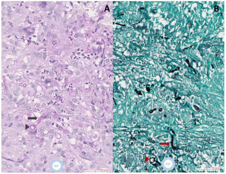
Figure 1.
Histopathological examination of skin biopsies demonstrating hyphal (arrows) and yeast structures (arrowheads) surrounded by epithelioid, multinuclear giant histiocytes and neutrophils in (A) Periodic acid–Schiff (black arrow and arrowhead), and (B) Grocott-Gomori (red arrow and arrowhead) stained slides, observed in 20× magnification.

