
Figure 3. Distinct and overlapping atrophy patterns in svPPA and bvFTD.
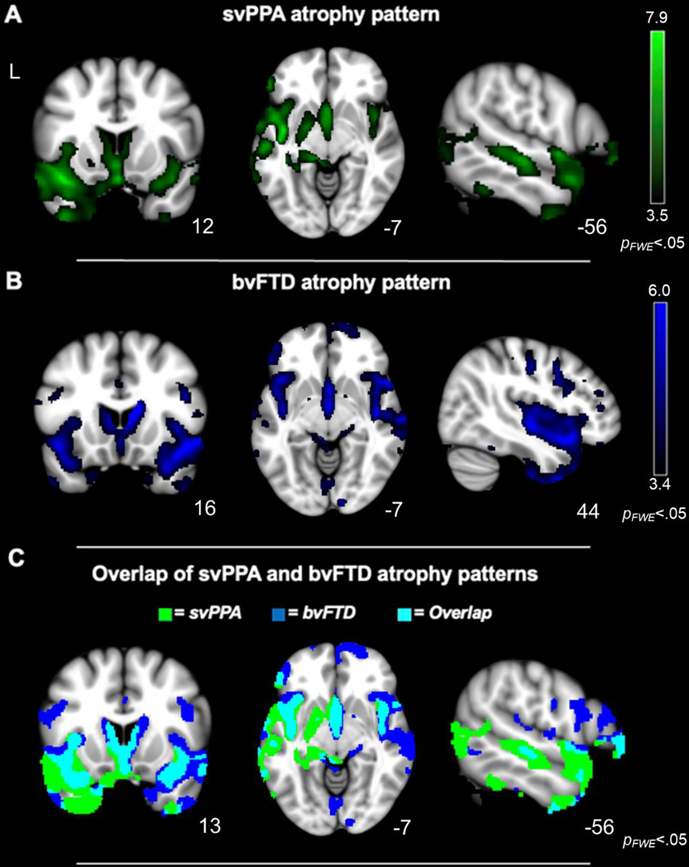
Whole-brain voxel-based morphometry analyses (controlling for age, sex, and TIV) confirmed that the A) svPPA and (B) bvFTD groups had smaller gray matter volume than the healthy controls in frontotemporal and subcortical regions typically atrophied in these syndromes (pFWE<.05). (C) In svPPA and bvFTD, there was some overlapping atrophy, but the people with svPPA tended to have more left lateralized atrophy than those with bvFTD, whose atrophy was more bilateral (pFWE<.05). Statistical maps are superimposed on the Montreal Neurological Institute template.