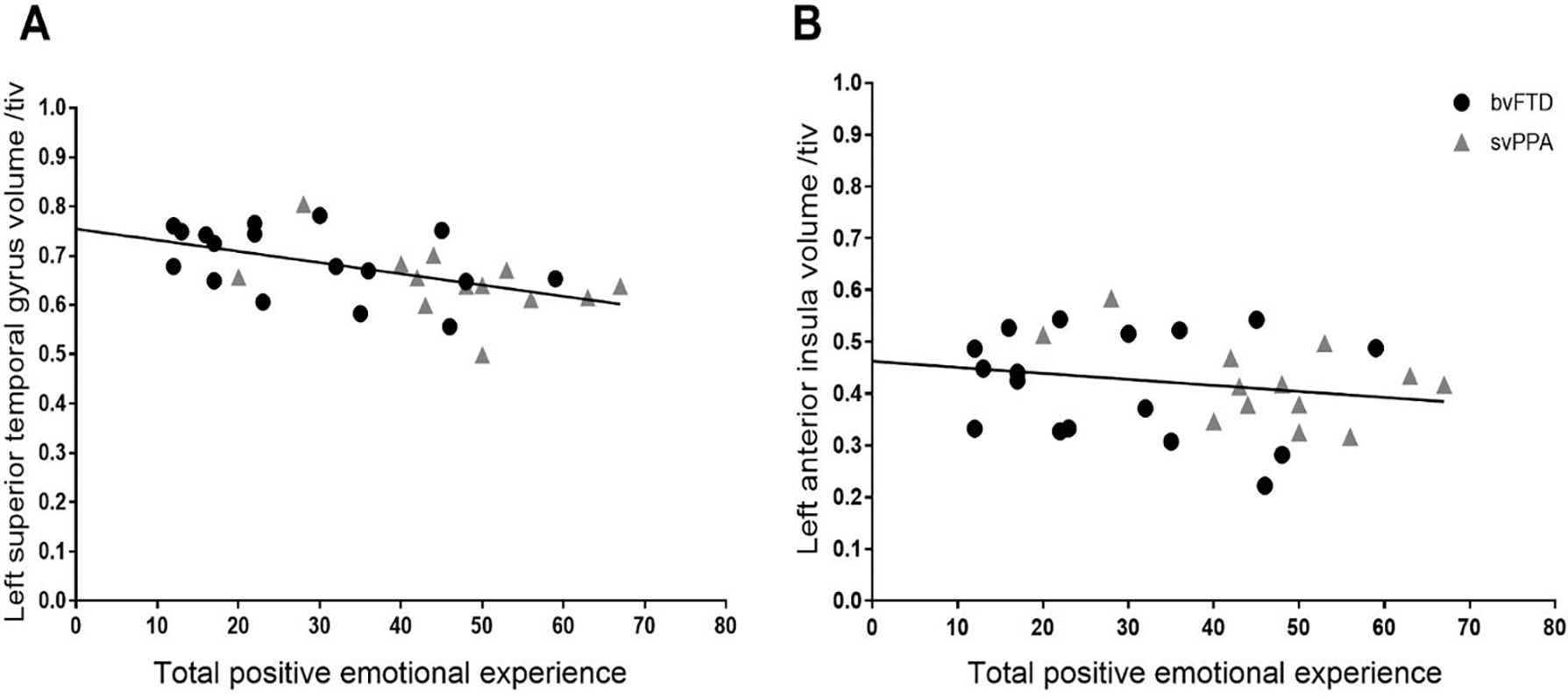
Figure 5. Linear association between total positive emotional experience and gray matter volume across the clinical groups.

A regression model (controlling for age, sex, diagnosis, TIV, and MMSE) confirmed that total positive emotional experience had a linear association across the clinical groups with gray matter volume in the left superior temporal gyrus and left dorsal anterior insula clusters that emerged in the VBM analyses. These results suggest the neuroimaging results reflected linear brain-emotion associations and were not driven by one diagnostic group.
