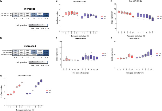
Fig. 6. Comparative analysis of time-course expression data show deregulation of microRNA expression in PD.
A, D Two core deregulated miRNAs with an adjusted p-value ≤ 0.05 and a median log2 FC ≥0.5 or ≤−0.5 for the comparison between PD and HC groups showed a significantly increased expression and three a significantly decreased expression at more than one time-point of the T cell activation course. Corresponding FDR adjusted p-values of the analyzed time-points (0, 2, 4, 8, 12, and 24 h) are depicted for the comparison between PD and HC groups. B, C and E–G Exemplary log2 time-course data are shown for hsa-miR-132-3p, hsa-miR-223-3p with an increased expression in PD and E–G for hsa-miR-4730, hsa-miR-762, hsa-miR-155-5p with a decreased expression in PD. The boxes show the range from the 25th to 75th percentiles with the median indicated by horizontal lines within the boxes and the total expression ranges per by whiskers. Significant differences between PD and HC at the time points are indicated by darker colors.

