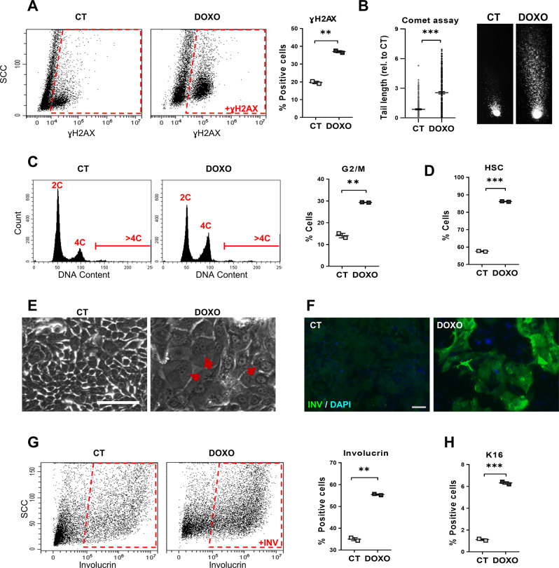
Fig. 1. Doxorubicin induces squamous metaplasia in human lung epithelial cells.
Human primary lung epithelial cells were treated with the dimethyl sulfoxide vehicle (CT) or with 0.5 μM Doxorubicin (DOXO) for 24 h (A, C) or 48 h (B, D–H). A Representative flow-cytometry analysis for the DNA damage marker γH2AX (+γH2AX, positive cells). Quantitation in the right histogram. B DNA fragmentation as analysed by comet assays, measured by tail length relative to CT (n = 245–247). Photographs show representative images of nuclei in CT or DOXO-treated cells as indicated. C Representative flow-cytometry analyses of DNA content of cells (2C, 4C, and >4C indicate diploid, mitotic/tetraploid and polyploid cells, respectively). Plot on the right shows percent of CT or DOXO cells in the G2/M phase of the cell cycle. D Percent of CT or DOXO-treated cells with high light scatter (HSC) typical of squamous differentiation, as analysed by flow cytometry. E Representative phase contrast images of cells treated for 48 h as indicated. Red arrows point at polyploid cells with several or large nuclei. Scale bar, 50 μm. F Immunofluorescence for the squamous differentiation marker involucrin (green). Blue is nuclear DNA by DAPI. Scale bar, 100 μm. G Representative flow cytometry analysis for involucrin (+INV, positive cells). The percent of involucrin positive CT or DOXO-treated cells is shown on the right. H Percent of squamous marker keratin K16 positive cells, as analysed by flow cytometry. Flow cytometry analysis gates were stablished according to negative isotype antibody control. Data are mean ± SEM of duplicate samples, representative of 2–3 independent experiment from two different human individuals with similar results. ***p ≤ 0.001, **p ≤ 0.01.

