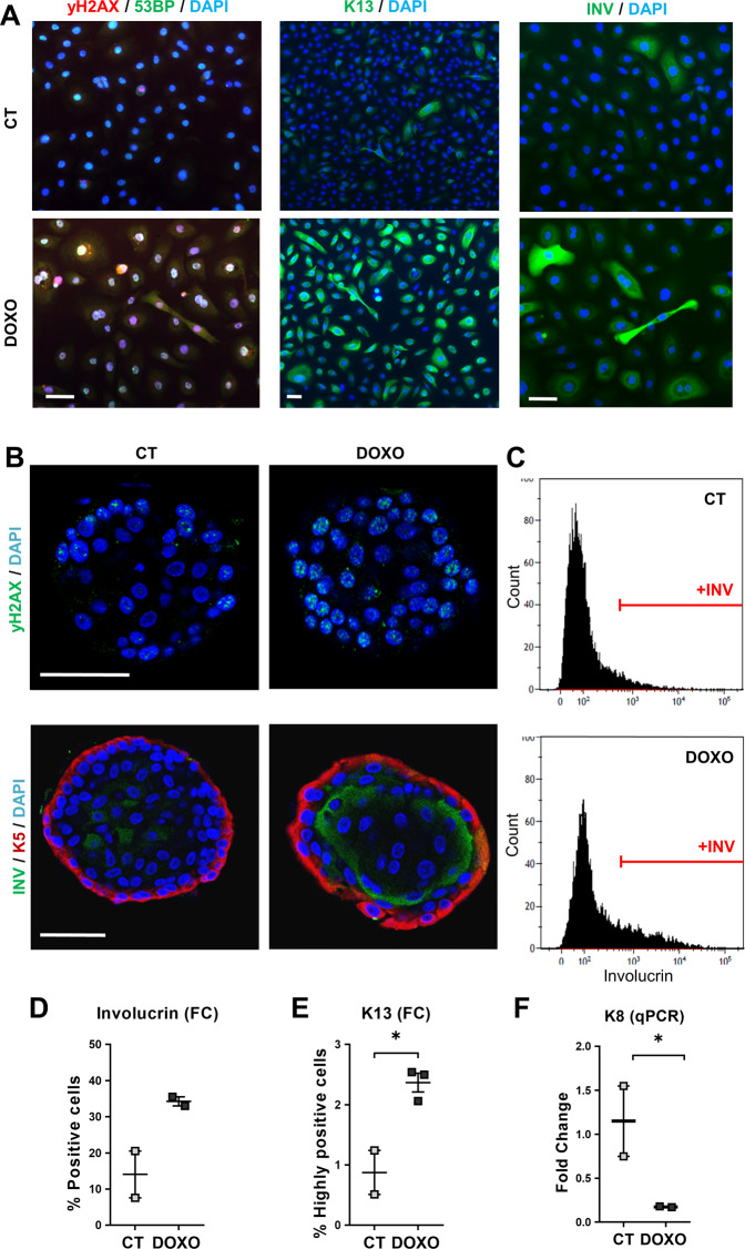
Fig. 2. Doxorubicin induces squamous differentiation in human lung epithelial cells in a serum-free lung-adapted medium and in organoid reconstructions.
A Human primary lung epithelial cells isolated and cultured in lung-adapted medium were treated for 48 h with the dimethyl sulfoxide vehicle (CT) or with 0.5 μM Doxorubicin (DOXO), as indicated. Immunofluorescence for 53BP (green) and γH2AX (red), for K13 (green), or for involucrin (green), as indicated. Blue is nuclear DNA by DAPI; scale bar, 50 μm. Representative of two independent experiments with similar results. B–F Airway lung organoids (AOs) in organoid expansion medium were treated with dimethyl sulfoxide (CT) or with 0.25 μM Doxorubicin (DOXO) for 24 h (C, D, F) or 48 h (B, E), as indicated. B Immunofluorescence confocal images of AOs treated as indicated and stained for γH2AX (top; green) or for involucrin (bottom; green) and K5 (red). Scale bar, 50 μm. Blue is nuclear DNA by DAPI. C Representative flow cytometry analyses for the differentiation marker involucrin (+INV, positive cells). D Percent of involucrin positive cells by flow cytometry. E Percent of keratin K13 positive cells by flow cytometry. F mRNA fold change of keratin K8 by qPCR. Positive cells by flow cytometry were gated according to negative isotype antibody control. Data are mean ± SEM of two or three replicate samples of two independent experiments from two different human individual. *p ≤ 0.05, p for +INV = 0.09.

