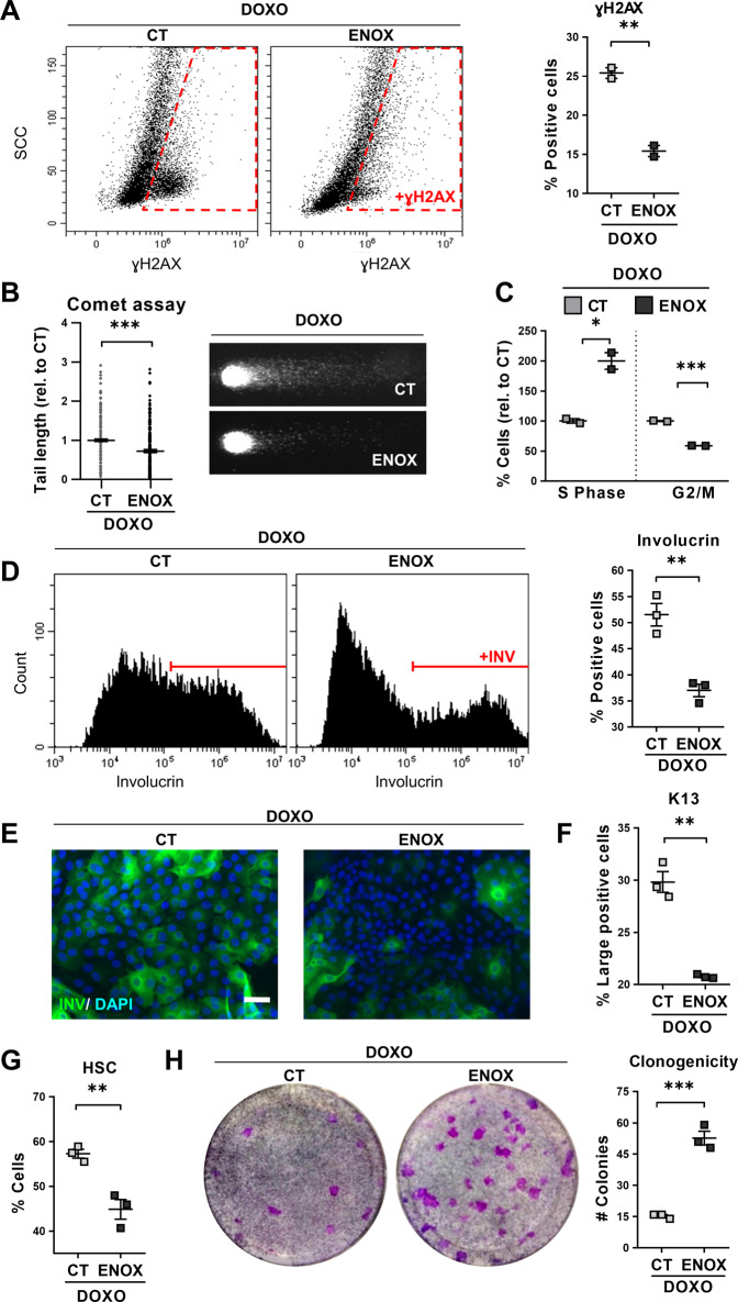
Fig. 5. Enhancement of DNA repair by Enoxacin suppresses Doxorubicin-induced squamous metaplasia in lung epithelial cells.
Human primary lung epithelial cells were treated with Doxorubicin (DOXO) and with the dimethyl sulfoxide vehicle (CT) or with 200 μM Enoxacin (ENOX). A Representative flow-cytometry analyses for the DNA damage marker γH2AX of cells treated for 24 h, as indicated (γH2AX+, positive cells). Scattered plot (right) shows percent of γH2AX positive cells. B DNA fragmentation as analysed by comet assays after 24 h treatment as indicated and measured by tail length relative to CT (n = 237–312). Photographs show representative images of nuclei in CT or ENOX-treated cells, as indicated. C Percent of cells in the S or G2/M phases of the cell cycle relative to CT, analysed by flow cytometry after 24 h with the indicated treatments. D Representative flow cytometry analyses for involucrin (+INV, positive cells) after 48 h with the indicated treatments. Positive cells were gated according to negative isotype antibody control. The percent of involucrin positive CT or ENOX-treated cells is shown on the right. E Immunofluorescence for involucrin (green) of cells treated for 48 h, as indicated. Blue is nuclear DNA by DAPI. Scale bar, 50 μm. F Percent of large keratin K13 positive cells analysed by flow cytometry after 24 h. G Percent of cells with high light scatter (HSC), analysed by flow cytometry after 24 h. H Clonogenic capacity of cells drug-released and plated after 48 h with the indicated treatments. The number of CT or ENOX colonies is shown on the right plot. Positive cells by flow cytometry were gated according to negative isotype antibody control. Data are mean ± SEM of 2–3 replicate samples, representative of 2–3 independent experiment from two different human individuals with similar results. ***p ≤ 0.001, **p ≤ 0.01, *p ≤ 0.05.

