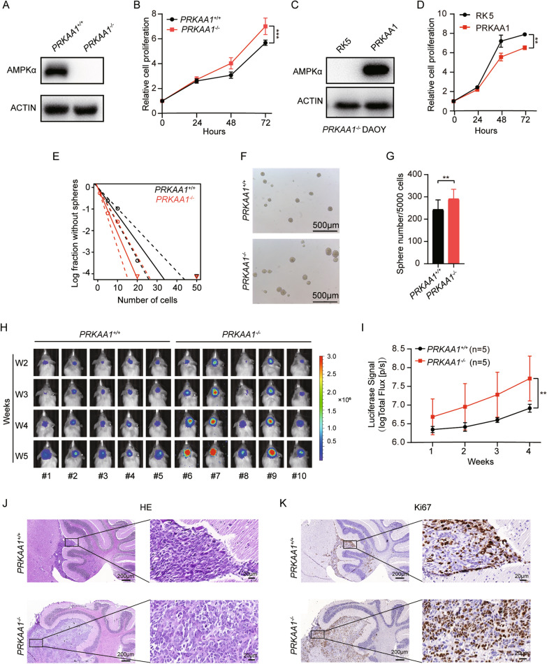
Fig. 2.
Knockout of AMPKα promotes DAOY proliferation. A Western blot detection of AMPKα protein level in PRKAA1+/+ and PRKAA1−/− DAOYs. B Proliferation of PRKAA1+/+ and PRKAA1−/− DAOYs were assessed by CCK-8. *** P < 0.001. C Western blot detection of AMPKα protein level in PRKAA1−/− DAOYs transfected with PRKAA1 cDNA. D Cell viability of transfected PRKAA1−/− DAOYs was detected by CCK-8. ** P < 0.01. E Tumorsphere formation ability was detected by extreme limiting dilution assays. F Representative images of tumorspheres formed by PRKAA1+/+ and PRKAA1.−/− DAOYs. G Quantification of the number of spheres (per 5000 cells) formed by DAOYs. Data in bar graphs are presented as mean ± SEM from three independent experiments. ** P < 0.01. H The time course of in vivo fluorescence images of NOD-SCID mice implanted with DAOY-Luc cells in the cerebellum using IVIS Spectrum In Vivo Imaging System. I Quantification of total Flux of mice. ** P < 0.01. J HE staining and K IHC staining of Ki67 of cerebellar tumors generated by implanting DAOY-Luc cells (5 × and 40x)

