Fig. 3.
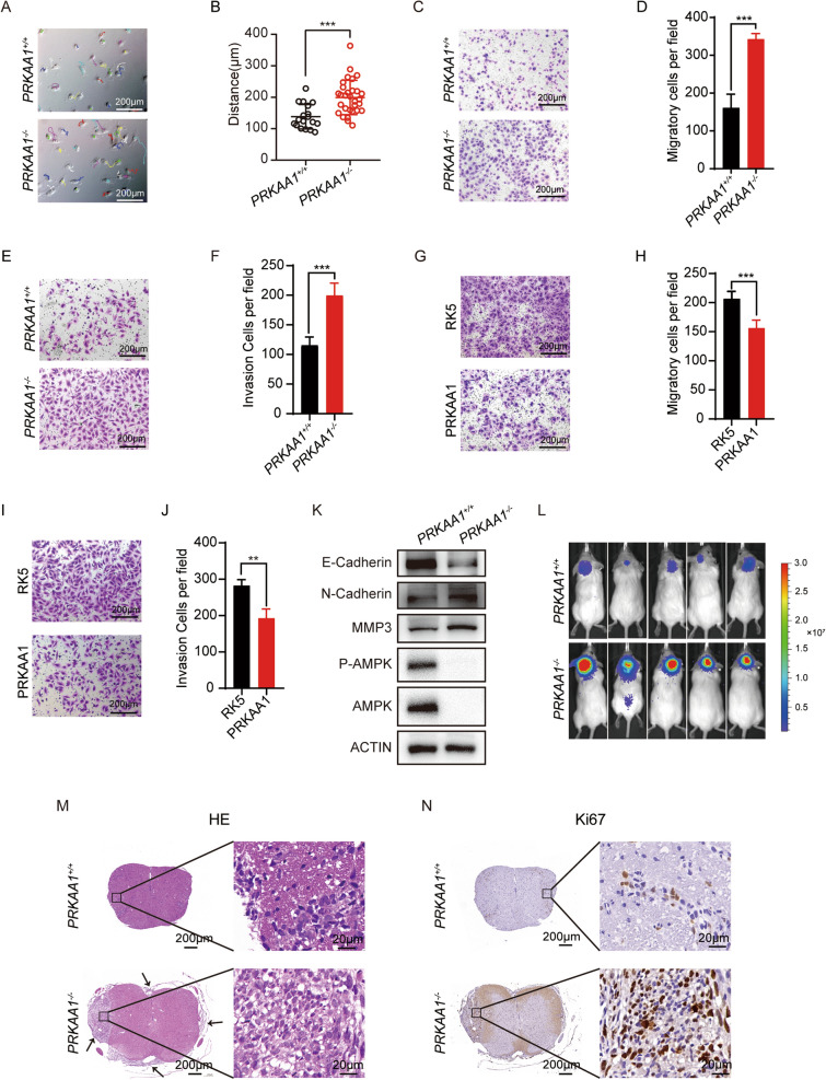
Knockout of AMPKα promotes DAOY migration and invasion. A-B Trajectory of DAOYs were recorded by Celldiscoverer 7 (Zeiss) and ImageJ. *** P < 0.001. C Transwell migration assay of knockout cells. D Quantification data performed the average migration ± SEM from three independent experiments. *** P < 0.001. E Transwell invasion assay of knockout cells with matrigel. F Quantification data performed the average invasion ± SEM from three independent experiments. *** P < 0.001. G and I Evaluation of migration and invasion ability of PRKAA1−/− DAOYs transfected with PRKAA1 plasmids by transwell assays with and without matrigel. H and J Quantitative data from three independent experiments performed as in G and I. ** P < 0.01. K Western blot detection of AMPKα, P- AMPKα and EMT markers in PRKAA1+/+ and PRKAA1.−/− DAOYs. L In vivo fluorescence images of NOD-SCID mice implanted with DAOY-Luc cells at Week7. M HE staining and N IHC staining of Ki67 of spinal cord from NOD-SCID mice implanted with DAOY-Luc cells (5 × and 40x)

