
Figure 1:
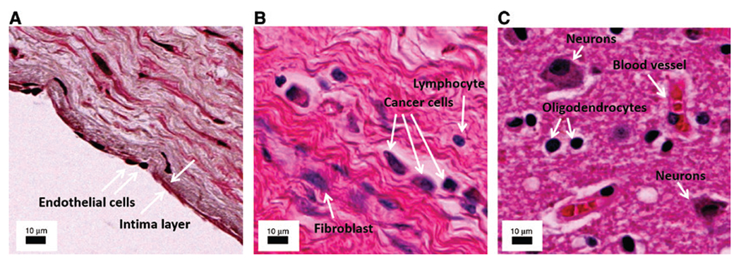
The need for single cell MSI.
The necessity for high resolution MSI becomes evident in these microscopic images of different clinical cases. (A) Human artery, arrows indicate one cell thick intima layer consisting of endothelial cells. (B) Human lobular breast carcinoma, arrows indicate cancer cells, a fibroblast, and a lymphocyte. (C) Human Alzheimer’s brain, arrows show the lymphocytes, an astrocyte, an oligodendrocyte, and a blood vessel. The annotations were performed to the best of our knowledge, however, additional cell-specific staining is necessary to confirm cell identities. The scale bar in all images is 10 μm.