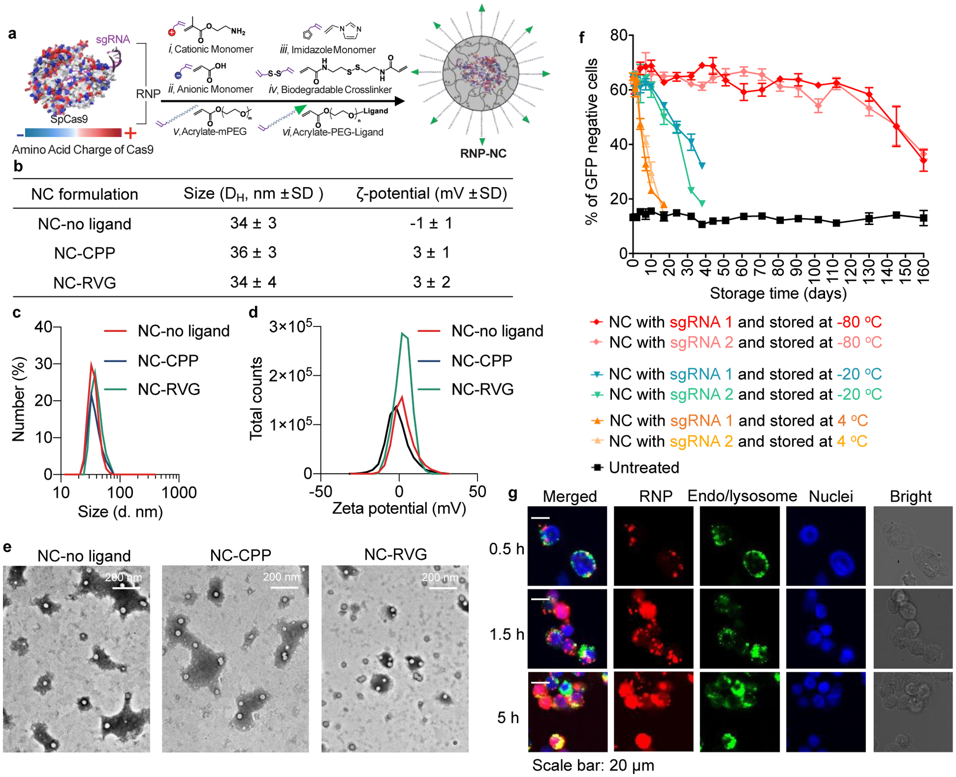
Figure 1. Synthesis and characterization of the RNP NCs.

a, A schematic illustration for the synthesis of RNP-encapsulated NC. b, Summary of the sizes and zeta-potentials of NCs with or without ligand. c, Size distribution of NC-no ligand, NC-CPP and NC-RVG. d, Zeta-potentials of NC-no ligand, NC-CPP and NC-RVG. e, TEM images of NC-No Ligand, NC-CPP and NC-RVG. f, RNP delivery of NC after storage at different conditions. g, Intracellular trafficking of NC-no ligand loaded with Atto550-tagged RNP. The intracellular localization of Atto550-tagged RNP was studied at 0.5 h, 1.5 h, and 5 h post-treatment. Intracellular trafficking of NC-CPP and NC-RVG are shown in Supplementary Figure 2. CPP, cell-penetrating peptide; GFP, green fluorescent protein; NC, nanocapsule; RNP, ribonucleoprotein; RVG, rabies virus glycoprotein; sgRNA, short guide RNA; TEM, transmission electron microscopy.
