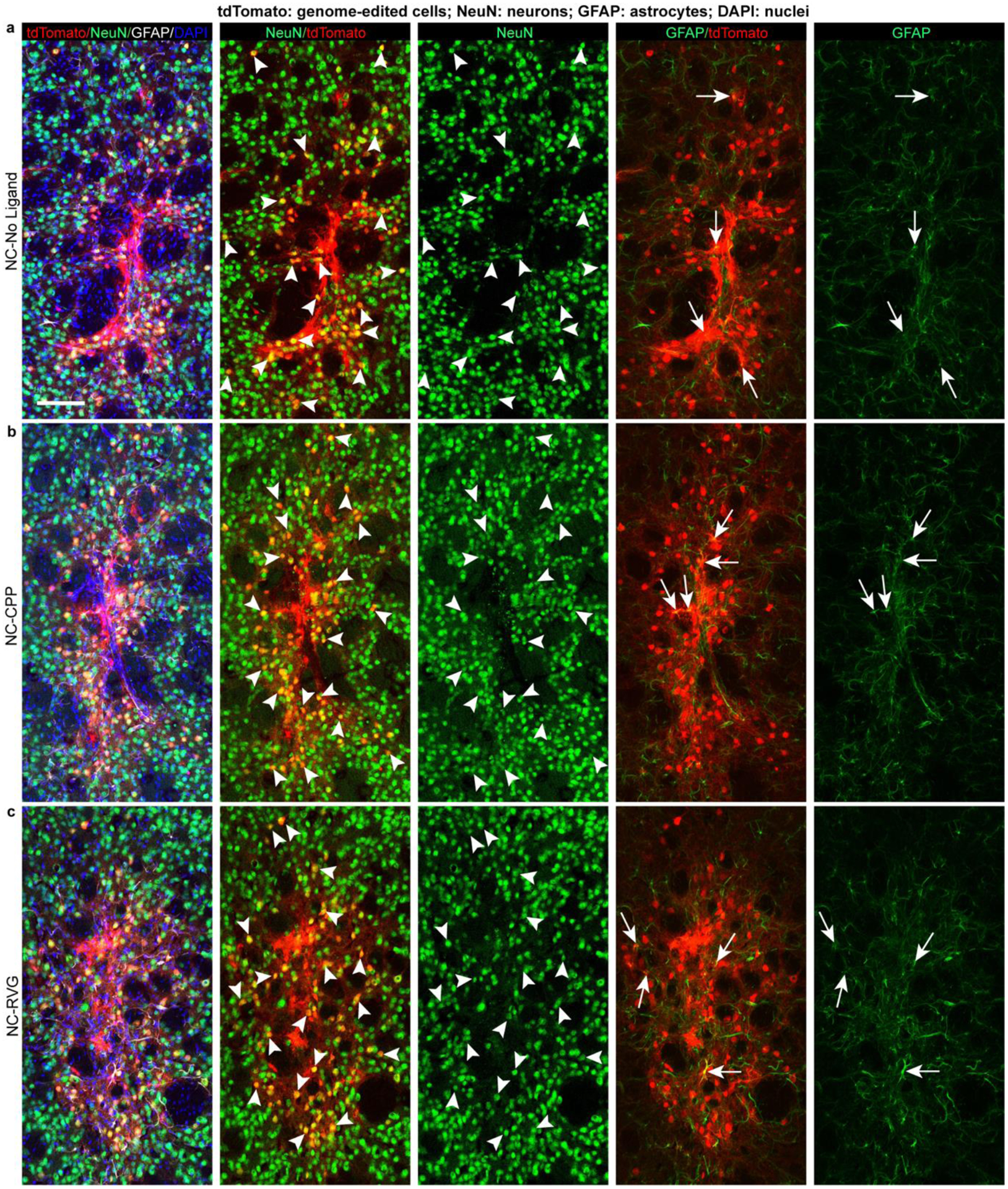
Figure 4. The majority of genome-edited cells following NC delivery are neuronal.

Genome-edited, tdTomato-expressing (red) cells in the striatum of representative animals from each of the NC treatment groups (NC-No Ligand animal J12; NC-CPP animal J18; NC-RVG animal J5; Supp. Table 1). Scale bar = 100 μm. Arrowheads point to examples of tdTomato+/NeuN+ genome-edited neurons (yellow in 2nd column). Arrows point to tdTomato+/GFAP+ genome-edited astrocytes (yellow in 4th column). Photomicrographs show maximum intensity projection of three focal planes covering 10 μm. Individual channels were adjusted for brightness as needed (Supp. Table 4). CPP, cell-penetrating peptide; DAPI, 4′,6-diamidino-2-phenylindole; GFAP, glial fibrillary acidic protein; NC, nanocapsule; NeuN, neuronal nuclear protein; RVG, rabies virus glycoprotein.
