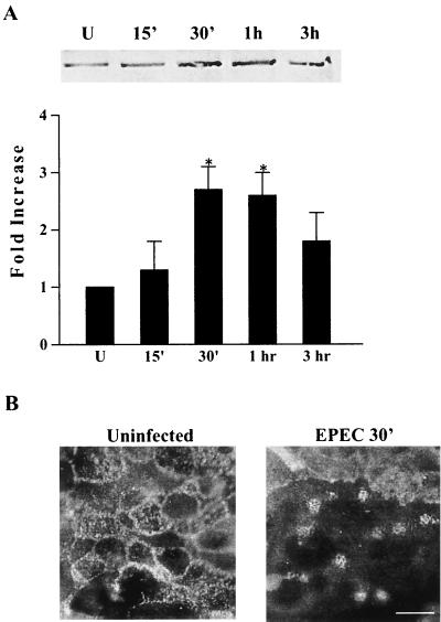
FIG. 2.
Threonine phosphorylation of ezrin is rapidly enhanced following EPEC infection and corresponds with A/E lesion formation. (A) Immunoprecipitation using antiphosphothreonine antibody was performed on extracts from uninfected controls (U) and cells infected with EPEC for the designated times. Separation by SDS-PAGE and immunoblot analysis using polyclonal ezrin antibody revealed an increase in ezrin threonine which peaked at 30 min postinfection. The bar graph represents means + standard errors of the means (error bars) of three separate experiments. *, P ≤ 0.05. (B) Uninfected monolayers and monolayers infected with EPEC for 30 min were subjected to immunofluorescence staining for ezrin. In uninfected controls, ezrin is distributed throughout the cells. After 30 min of EPEC infection, characteristic A/E lesions have formed and ezrin redistribution to these sites is evident. Bar,10 μm.

