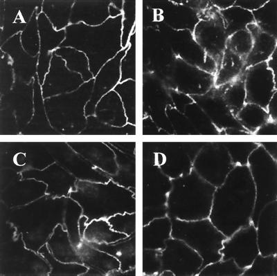
FIG. 8.
Dominant-negative ezrin diminishes the effect of EPEC on ZO-1. Confluent monolayers of E17 and N12 cells were subjected to immunofluorescence staining for ZO-1. Uniform distribution of ZO-1 at the level of TJ was observed in both E17 (A) and N12 (C) monolayers. (B) After 3 h of EPEC infection, breaks in the staining of ZO-1 were seen in E17 cells. (D) The alterations in ZO-1 staining in response to EPEC infection were markedly attenuated in N12 monolayers expressing dominant-negative ezrin.

