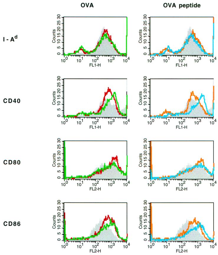
FIG. 6.
Mφ incubated with CTB-coupled antigens up-regulate their cell surface expression of CD40 and CD86 in vitro. Mφ were pulsed with OVA, CTB-OVA, OVA peptide, or CTB-coupled OVA peptide, or were left untreated, and then incubated for 24 h with OVA-specific TCR-transgenic T cells. The Mφ cell surface levels of MHC II, CD40, CD80, and CD86 were then evaluated by FACS analysis. Data are presented as fluorescent intensities and were recorded for unpulsed (shaded), OVA-pulsed (red line), and CTB-OVA-pulsed (green line) Mφ (A) and for unpulsed (shaded), OVA peptide-pulsed (orange line), and CTB-OVA peptide-pulsed (blue line) Mφ.

