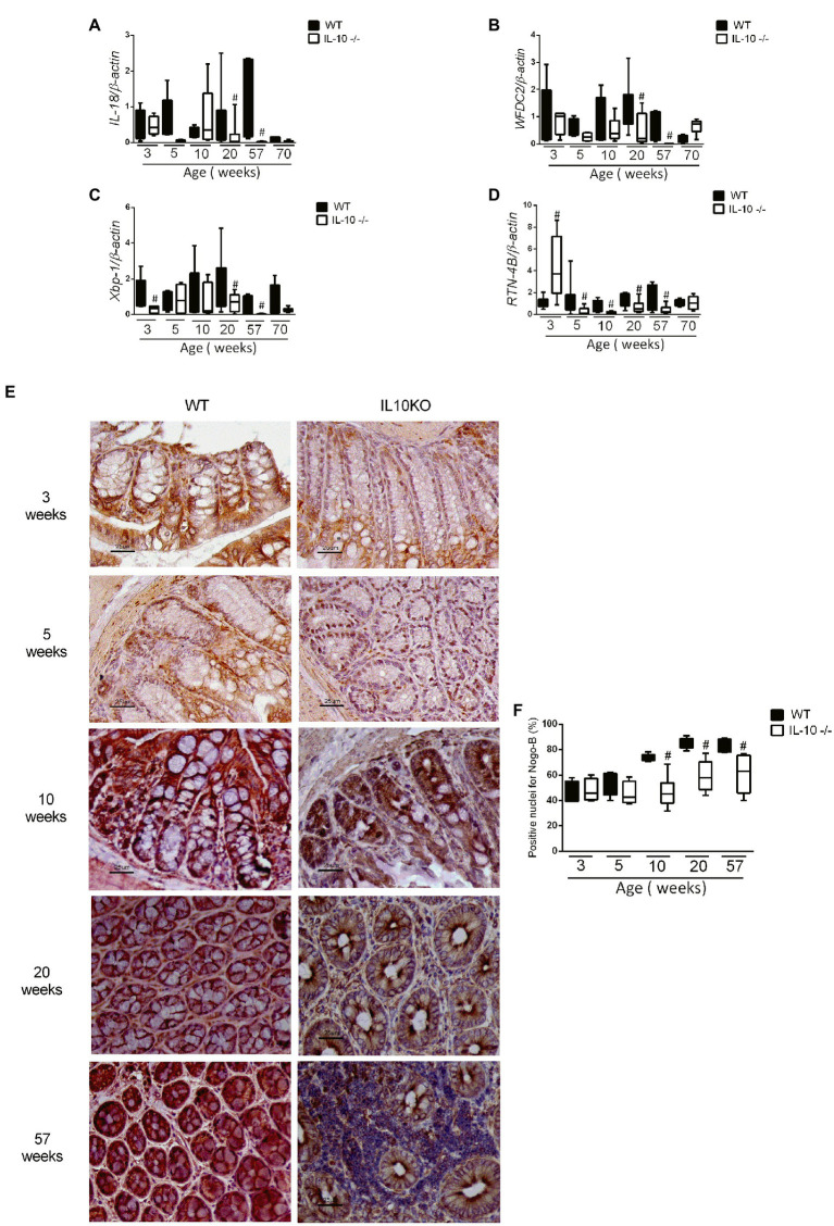
Figure 5.
Analysis of gene expression by qPCR of IL-18 (A), WFDC2 (B), Xbp1 (C), and RTN-4B/NogoB (D) in the colon of WT and KO mice in all evaluated ages. C57Bl/6 wild type (WT) group was represented in black and IL-10 deficient mice (IL-10−/−) group in white. #p < 0.05 with respect to WT of the same age. (E) Histological sections of the colon of WT and IL-10−/− mice at 3, 5, 10, 20, and 57 weeks of age labeled with RTN-4B antibody and revealed with DAB (n = 6 for each strain and age). (F) RTN-4B positive nuclei count from images acquired in ACT-1 software in blindly and independent manner.

