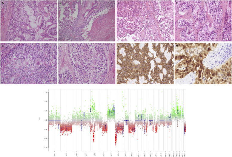
Figure 2.
Histological and mutational characteristics of pleural disease. Histologically, this tumor comprises a mixture of papillary, solid and glandular architectures (A–D). The cells are arranged around variably well-formed fibrovascular cores (A and B), with prominent areas of myxoid stroma (A and C). Focal tumor necrosis is present (B). At higher magnification (D) the tumor is composed of cells with relatively small, bland, ovoid, vesicular nuclei with abundant fibrillary or focally clear cytoplasm. No pleomorphism or mitotic activity is discernible (hematoxylin and eosin). At low power (E), there is prominent papillary architecture with stromal cores being partly myxoid. A separate area (F) shows organoid nests of mildly atypical cells. Tumor cells stain diffusely for CD56 (G). Most tumor cells stain diffusely for S-100 (G). M-ethylation array profiling of pleural tissue (I) using the Illumina Infinium 850k EPIC array analysed as per the DKFZ Heidelberg algorithm, confirming myxopapillary ependymoma (MPE).

