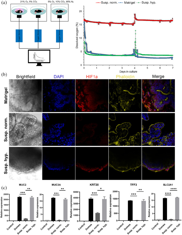
Figure 2.
HIF-1α-induced effects on apical-out small intestinal organoids. (a) Sensor measurements of oxygen levels in organoids cultured in Matrigel domes, suspension normoxia, and suspension hypoxia over a period of 7 days. For organoids grown in suspension normoxia, the dissolved oxygen concentration was about 15%–18%, whereas in Matrigel-embedded and suspension hypoxia cultures, the dissolved oxygen concentration was about 3%–5%. The peak at day 4 corresponds to medium refreshment. Error bars indicate mean ± S.E.M. (n = 3). (b) Immunofluorescence stainings indicated the expression of HIF-1α in low oxygen conditions (Matrigel-embedded and hypoxia suspension). No expression was identified in organoids grown in suspension normoxia. (c) Comparison of gene expression levels of HIF-1α targets, including MUC2, MUC3A, KRT20, TFF3, and SLC2A1 between the three culture conditions. All these genes were significantly upregulated in low oxygen conditions. Error bars indicate mean ± S.E.M. (n = 3).

