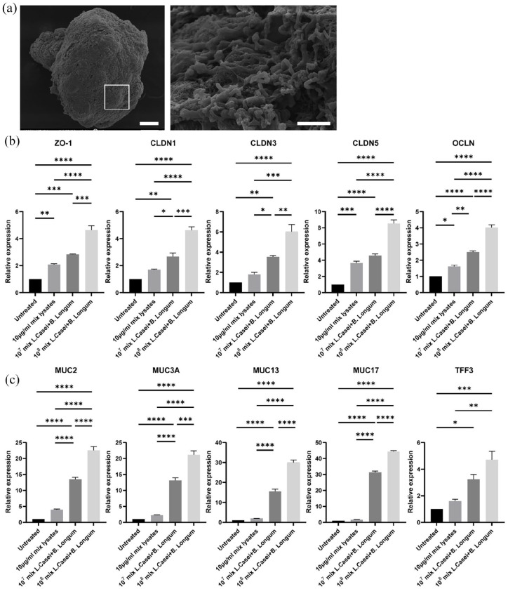
Figure 7.
Triple co-culture of organoids with L. casei and B. longum. (a) SEM microscopy indicated colonization of L. casei and B. longum bacteria on the apical surface of the organoids. The white box in the left image represents the area magnified in the corresponding image on the right. Scale bars: 100and 5 μm. (b and c) qRT-PCR analysis demonstrated the expression levels of the junction markers ZO-1, OCLN, CLDN1, CLDN3, and CLDN5 (b) and the mucins markers MUC2, MUC3A, MUC13, MUC17, and TFF3 (c) in organoids co-cultured with a 50:50 mix of L.casei- and B.longum-derived lysates (final concentration 10 μg/ml) and a 50:50 mix of L.casei and B. longum cells (final concentrations: 107 and 108). Error bars indicate mean ± S.E.M. (n = 3).

