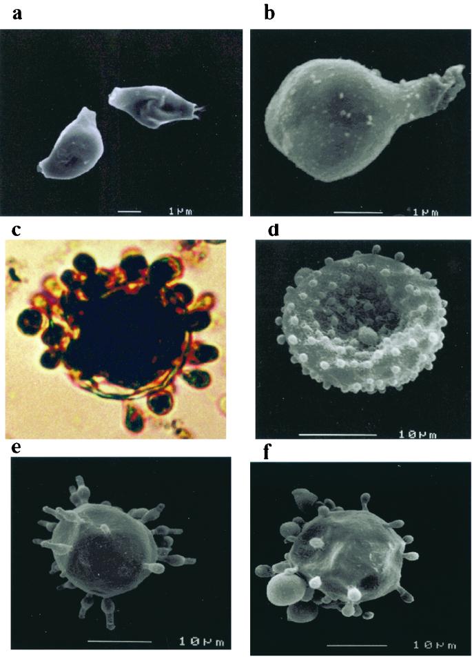
FIG. 1.
Scanning electron micrographs of P. brasiliensis conidia and photomicrographs and scanning electron micrographs of yeast cells (ATCC 60855) before and after treatment with proteinases, guanadinium isothiocyanate, and hydrochloric acid. (a and b) Conidia before and after treatment respectively, (c and d) light photomicrograph and scanning electron micrograph, respectively, of P. brasiliensis (ATCC 60855) yeasts grown on l-DOPA (without treatment); (e and f) yeast cells grown on minimal media supplemented with l-DOPA before and after treatment, respectively.

