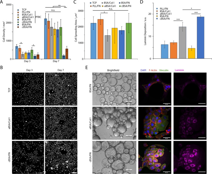
Figure 4.
(A) HPK proliferation on interfaces conditioned with different supercharged albumins, functionalized with ECM proteins and assembled with or without co-surfactant PFBC. (B) Selected images of cells spreading at corresponding liquid–liquid interfaces after 3 and 7 days of culture at TCP, cBSA/FN, and aBSA/Col I with PFBC. Images are corresponding nuclear stainings. Scale bars are 200 μm. (C) Quantification of HPK spreading area (24 h after seeding) characterized on pinned droplets functionalized with corresponding nanosheets. (D) Quantification of laminin deposition at liquid–liquid interfaces. (E) Bright-field and confocal images of HPKs cultured for 7 days on emulsions stabilized by protein nanosheets (blue, DAPI; red, phalloidin; green, vinculin; and purple, laminin). Scale bars are 100 μm (bright-field) and 50 μm (confocal). Error bars are s.e.m.; n = 3.

