FIGURE 1.
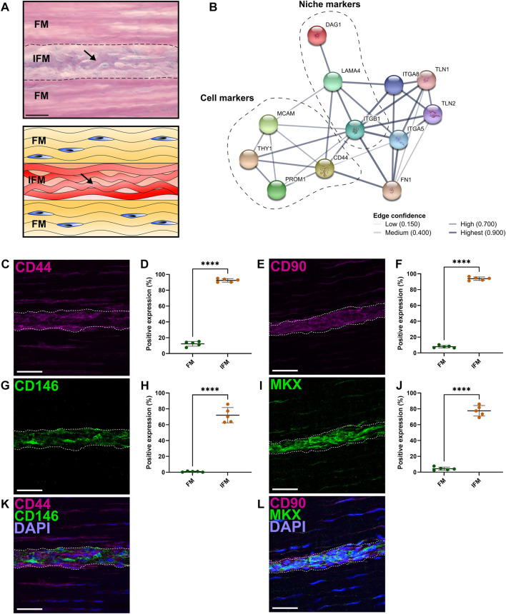
Analyses of regional differences in tendon cell marker expression demonstrated that CD146 is exclusively expressed by interfascicular cells within an interfascicular niche. (A) PAS-staining and schematic of SDFT sections highlighted mucin-rich basement membrane (arrow; purple, schematic; red) within the interfascicular matrix (IFM). Nuclei = blue. Scale bar = 50 µm. (B) STRING-predicted protein-protein interactions revealed potential targets for novel tendon cell populations using validated interfascicular niche markers CD146 and LAMA4. Interactions based on CD146 (MCAM) and LAMA4 demonstrated a protein neighborhood consisting of cell markers CD44, CD90 (THY1), CD133 (PROM1), as well as cell niche components such as ITGB1, DAG1 and FN1. (C–J) Image analyses comparing the positive labelling (area fraction; %) of longitudinal SDFT sections immunolabelled with CD44 (C,D), CD90 (E,F), CD146 (G,H) and MKX (I,J) overlayed with DAPI [blue = nuclei; (K,L)] in both fascicular matrix (FM) and IFM regions. The IFM is outlined by dotted lines. Scale bar = 50 µm. Biological replicates (n) = 5 per tendon region. Technical replicates = 3-4 per individual sample. Graphs were plotted as mean (µ) ± SD. Statistical significance: **** (p ≤ .0001).

