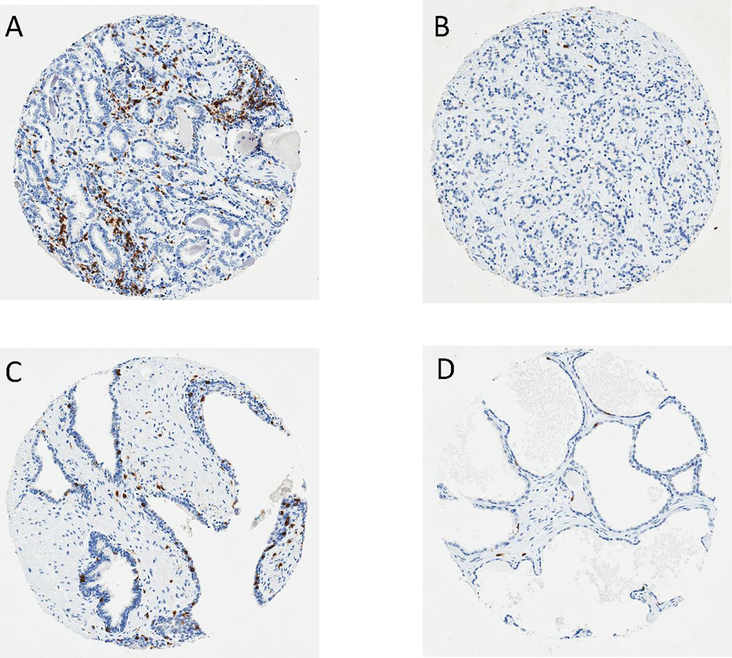
Figure 1. CD8 immunohistochemical staining of primary prostate cancer tissue microarrays.

Representative images (100 X) of CD8 immunostaining (in brown) show (A) Tumor core with high CD8+ TIL density; (B) Tumor core with low CD8+ TIL density; (C) Benign core with high CD8+ TIL density; (D) Benign core with low CD8+ TIL density.
