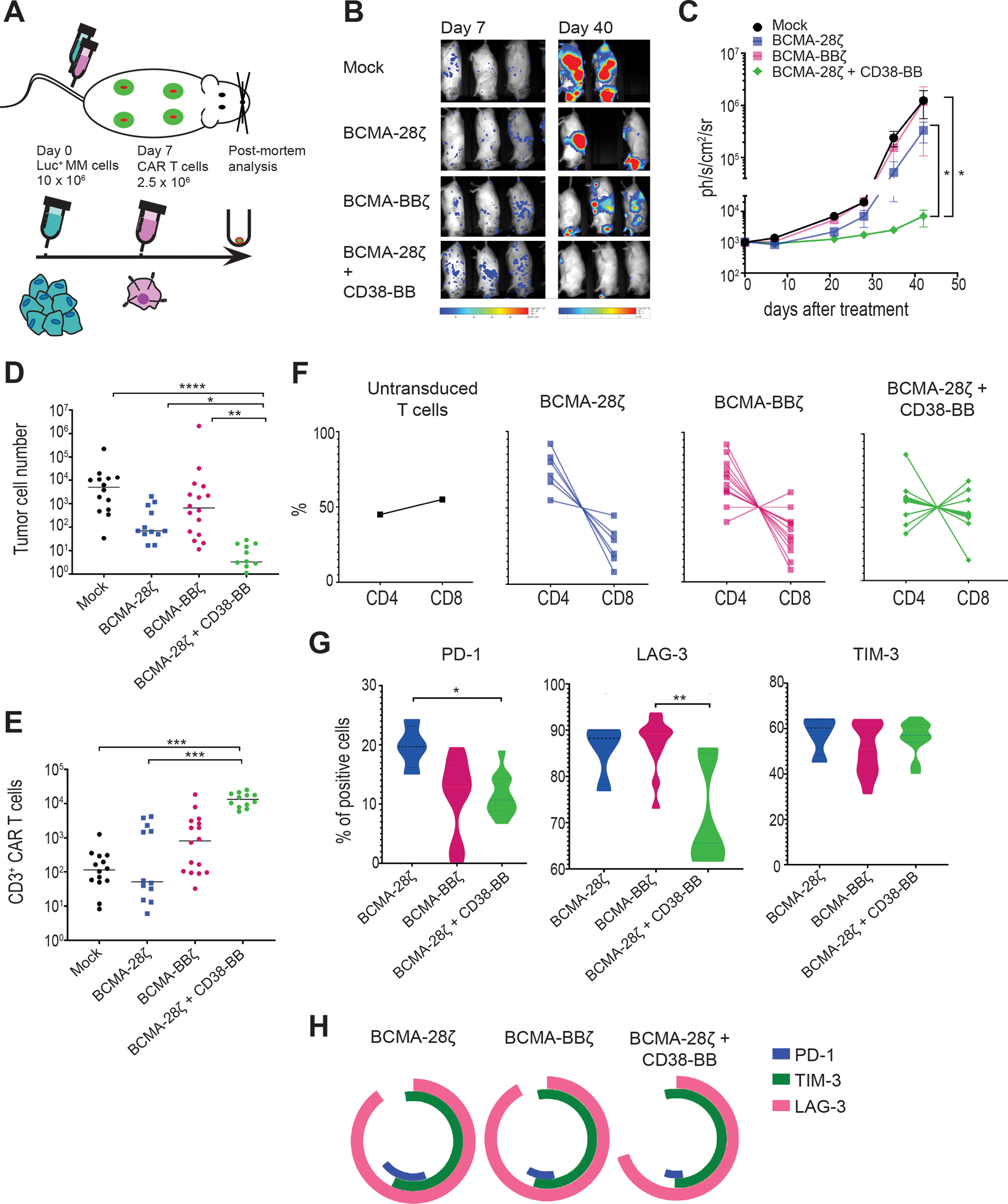
Figure 6. BCMA-CAR+CD38-CCR T cells show enhanced in vivo anti-tumor function, improved persistence, and reduced expression of exhaustion markers.

(A) A schematic representation of the MM scaffold-based xenograft murine model is shown. 10 × 106 Luc-GFP UM9 cells were administrated intravenously. Mock-transduced, BCMA-28ζ, BCMA-BBζ or BCMA-28ζ+CD38-BB CAR T cells were infused intravenously 7 days later. Tumor burden was measured weekly by BLI. (B) Representative BLI images of two time points are shown, with the pixel intensity represented in color. (C) Average tumor burden of mice was quantified by BLI and is depicted as units of photons per second per square centimeter per steradian (ph/sec/cm2/sr) (n=4 mice per group). Statistical analysis was performed using a two-way ANOVA and subsequent multiple comparison, corrected by Turkey test. *p<0.05. (D to H) Post-mortem scaffolds were harvested from each mouse and dissociated. Single-cell suspensions were counted, stained, and measured by flow cytometry. (D) Absolute UM9 tumor cell (GFP+/CD38+) numbers in the scaffolds are shown. Each dot represents one scaffold. (E) Absolute CAR T cell numbers in the scaffolds are shown. Each dot represents one scaffold. (F) CD4 and CD8 CAR T cell percentages in each scaffold are shown. (G) Violin plots show the expression of PD-1, LAG-3, and TIM-3 on CAR T cells isolated from scaffolds. (H) Co-expression of inhibitory receptors of (G) are presented. Statistical comparisons in (D), (E), and (G) were performed by Kruskal-Wallis test between the indicated groups; ns, not significant. *p<0.05, **p<0.01, ***p<0.001, ****p<0.0001.
