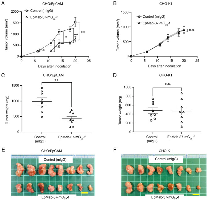
Figure 3.
Antitumor activity of EpMab-37-mG2a-f. (A and B) Measurement of tumor volume in (A) CHO/EpCAM and (B) CHO-K1 xenograft models. CHO/EpCAM and CHO-K1 cells (5×10⁶ cells) were injected into mice subcutaneously. On day 6, 100 µg EpMab-37-mG2a-f or mIgG were injected into mice intraperitoneally. On day 14, additional antibodies were injected. On days 6, 11, 14, 18 and 20 following the inoculation, the tumor volume was measured. Values are presented as the mean ± SEM. *P<0.05 and **P<0.01 (ANOVA and Sidak's multiple comparisons test). (C and D) The weight of the excised (C) CHO/EpCAM and (D) CHO-K1 xenografts was measured on day 20. Values are presented as the mean ± SEM. **P<0.01 (Welch's t-test). (E and F) The resected tumors appearance of (E) CHO/EpCAM and (F) CHO-K1 xenografts in the control mouse IgG and EpMab-37-mG2a-f treated groups on day 20 (scale bar, 1 cm). n.s., not significant; CHO, Chinese hamster ovary; EpCAM, epithelial cell adhesion molecule; mIgG, mouse IgG.

