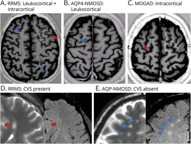
Figure 2. Examples of Cortical Lesions Seen on Phase-Sensitive Inversion Recovery (PSIR) Images and Lesions With and Without the Central Vein Sign (CVS) on Susceptibility-Weighted Imaging (SWI) in RRMS, AQP4-NMOSD, and MOGAD.
In the upper figures, PSIR imaging showing lesions located exclusively in the cortex (intracortical, red arrow) or within the cortex and adjacent juxtacortical white matter (leukocortical, blue arrow) in RRMS, AQP4-NMOSD, and MOGAD. Intracortical and leukocortical lesions were detected in patients with RRMS (A), whereas 1 leukocortical lesion in 1 patient with AQP4-NMOSD (B) and 1 intracortical lesion in 1 patient with MOGAD (C) were found. In the lower figures, T2 and corresponding SWI of deep white matter lesions with (red arrow) or without (blue arrow) CVS in RRMS and AQP4-NMOSD. The dark vein was located centrally in a lesion in an RRMS patient (D), while it was absent in three lesions in an AQP4-NMOSD patient (E). AQP4-NMOSD = aquaporin-4 antibody–positive neuromyelitis optica spectrum disorder; MOGAD = myelin oligodendrocyte glycoprotein antibody–associated disease; RRMS = relapsing-remitting multiple sclerosis.

