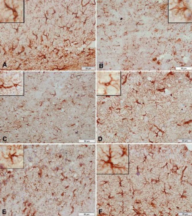
Figure 11.
Representative photomicrographs of cerebral cortex sections stained immunohistochemically with GFAP antibody in rats treated with: (A) Vehicle: the positive reaction of glial cells in the normal cerebral cortex. (B) Rotenone: marked decrease in positive reaction. Notice the decrease in cell body size of glial cells. (C) Rotenone+rasagiline: mild increase in positively stained glial cells. (D) Rotenone+L-dopa: mild increase in positively stained glial cells. (E) Rotenone+L-dopa+curcumin: noticeable increase in positively stained cells, though they still have small bodies and short dendrites. (F) Rotenone+rasagiline+curcumin: positive reaction close to normal. Note the large cell body and long dendrites of glial cells in the upper left part of the figure

