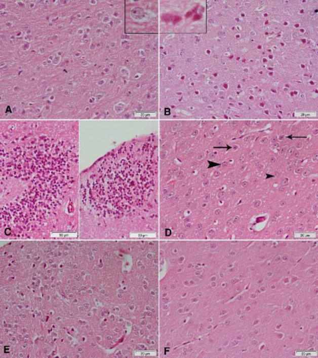
Figure 8.
Representative photomicrographs of cerebral cortex sections stained with Hx & E
(A) Vehicle: normal structure of neurons with characteristic large vesicular nuclei. (B) Rotenone: many neurons appear smaller than normal and deeply stained. The upper left part of the figure shows neurons with eosinophilic cytoplasm and pyknotic or fragmented nuclei. (C) Rotenone: focal infiltration with inflammatory cells. (D) Rotenone and rasagiline: amelioration of the damaging effect of rotenone, though some small dark neurons (arrows) and karyorrhectic (arrowheads) are still observed. (E) Rotenone and curcumin: most of the neurons appear normal with few small dark neurons. (F) Rotenone+rasagiline+curcumin: quite normal cerebral cortex tissue

