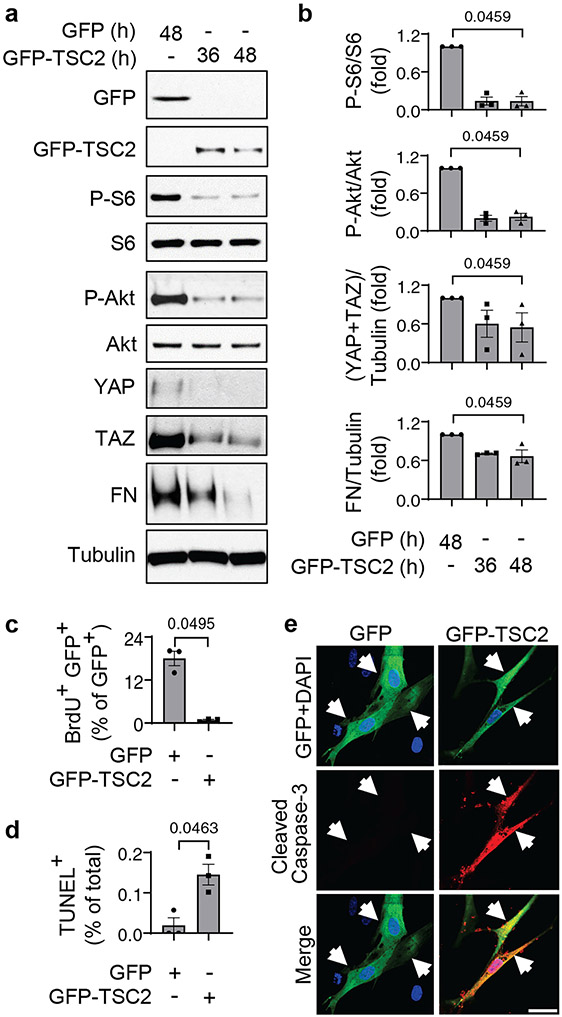
Figure 4. TSC2 reconstitution reduces YAP/TAZ accumulation and fibronectin production, suppresses proliferation, and induces apoptosis in human PAH PAVSMCs.
a, b. Human PAH PAVSMCs were transfected with GFP or GFP-TSC2 for the indicated times and immunoblotted to detect the indicated proteins. Data are means±SE from 3 subjects/group. P values for GFP-TSC2 compared to GFP were determined by Kruskal-Wallis rank test with Dunn’s pairwise comparison.
c-e. Proliferation (as measured BrdU incorporation) (c), apoptosis (as measured by TUNEL staining) (d), and immunocytochemical analysis to detect cleaved caspase 3 (red), GFP (green), and DAPI (blue) (e). Data are means±SE from 3 subjects/group. A minimum of 12 transfected cells/subject and condition were analyzed. P values for GFP-TSC2 compared to GFP were determined by Mann Whitney U test. Scale bar, 50 μm. White arrows indicate transfected cells.

