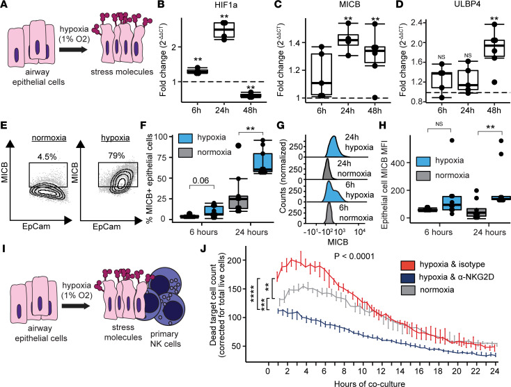
Figure 5. NKG2D stress ligands mediate killing of airway epithelial cells. (A).
Airway epithelial cells grown in liquid culture were subjected to hypoxia (1% O2) or normoxia (21% O2). (B–D) Transcription at 6 hours (6h), 24 hours (24h), and 48 hours (48h) was measured by PCR for hypoxia inducible factor 1A (HIF1A), MICB, and ULBP4. (E) Representative flow cytometry plots of surface MICB with 24 hours of normoxia or hypoxia. (F) Airway epithelial cell surface expression of MICB. (G) Representative histograms of MICB median fluorescence (MFI). (H) MICB MFI is shown at 6 and 24 hours. (I) To assess NK cell killing of airway epithelial cells, hypoxic or normoxic control cells were cocultured in a 2:1 ratio for 24 hours with primary human NK cells. Hypoxic cells were treated with isotype-matched control antibody or anti-NKG2D blocking antibody for 24 hours preceding the experiment. (J) Dead cell counts are shown at 30-minute intervals across 24 hours comparing total AUC for the 3 conditions. Summary data are displayed with box-and-whisker plots illustrating individual data points, bounded by boxes at 25th and 75th percentiles, and with medians depicted with bisecting lines. Individual P values calculated with Mann Whitney U test (B, C, D, F, and H) and 1-way ANOVA with Tukey’s honestly significant difference post hoc comparisons. ** P < 0.01, *** P < 0.001, **** P < 0.0001.

