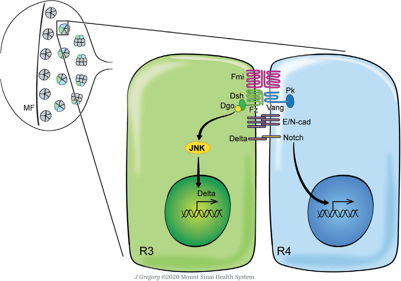
Fig. 2.

Planar cell polarity signaling and ommatidial rotation in the Drosophila eye. The Drosophila eye develops from an epithelial imaginal disc during larval stages, which is initially composed of identical pluripotent precursor cells ahead of the morphogenetic furrow (MF) (upper left panel). As the MF sweeps across the disc from posterior to anterior, preclusters of differentiating cells start to emerge which will then develop into mature ommatidia (Cagan & Ready, 1989; Roignant & Treisman, 2009; Tomlinson & Ready, 1987). During differentiation and maturation, these clusters rotate 90° in opposite directions in the dorsal and ventral halves of the eye to establish the final mirror-symmetric pattern of ommatidia across the D/V midline ( Jenny, 2010; Mlodzik, 1999). Posterior to the MF, Fz/PCP signaling mainly takes place between the photoreceptors R3 (green) and R4 (blue), inducing R4 fate via Notch signaling activation (Cooper & Bray, 1999; Das, Jenny, Klein, Eaton, & Mlodzik, 2004; Das, Reynolds-Kenneally, & Mlodzik, 2002; del Alamo & Mlodzik, 2006; Fanto & Mlodzik, 1999; Tomlinson & Struhl, 1999; Wolff & Rubin, 1998; Wu, Klein, & Mlodzik, 2004). The core PCP factors become asymmetrically localized in the subapical membrane region of the R3/R4 pair cell boundary, leading to differential downstream nuclear and/or cellular responses in each cell mediated by signaling (JNK, Delta, Notch) and simultaneously adhesion (E- and N-cadherin) molecules which are thought to mediate the cell motility process of ommatidial rotation (Cooper & Bray, 1999; Das et al., 2002, 2004; del Alamo & Mlodzik, 2006; Fanto & Mlodzik, 1999; Mirkovic et al., 2011; Mirkovic & Mlodzik, 2006; Tomlinson & Struhl, 1999; Wolff & Rubin, 1998; Wu et al., 2004). Dorsal is up, anterior is left. See main text for details.
