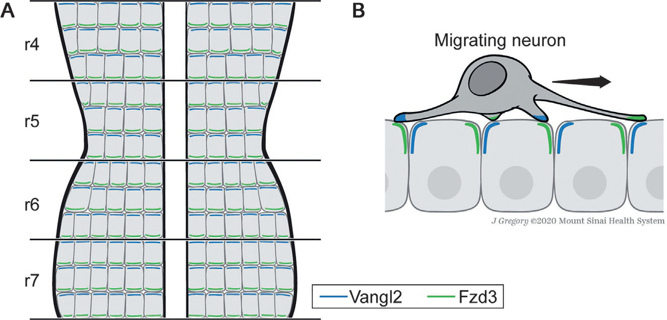
Fig. 5.

Model of PCP-mediated FBMN migration in the vertebrate hindbrain. The vertebrate hindbrain is segmented into developmental units called rhombomeres during embryonic development. FBMNs are born in rhombomere 4 (r4) and tangentially migrate to the more posterior r7 (Chandrasekhar, 2004). In zebrafish, rhombomeres have been shown to be planar polarized with Fzd3 and Vangl2 being asymmetrically enriched in anterior and posterior (sub)apical membranes, respectively (A) (Davey, Mathewson, & Moens, 2016). Migrating FBMNs enrich filopodial protrusions over the neuroepithelium in the direction of migration. Vangl2 becomes transiently enriched at the tips of filopodia in FBMNs preceding retraction, suggesting that transient PCP-mediated signaling events between FBMNs and the polarized neuroepithelium may promote FBMN migration (B) (Davey et al., 2016). Anterior is up. See text for details.
