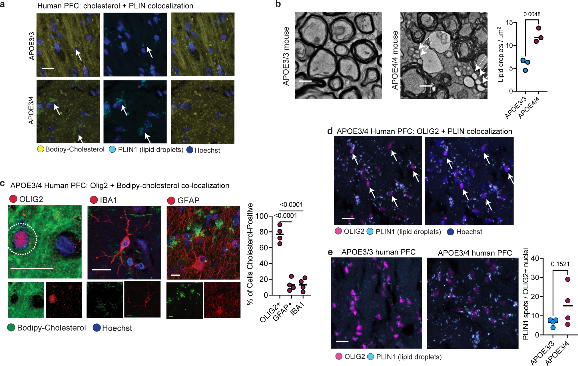
Extended Data Fig. 4: Lipid droplets in human and mouse brain.

a, Co-localization of immunohistochemistry against lipid-droplet associated protein perilipin-1 (PLIN1) with Bodipy-cholesterol staining in the human prefrontal cortex from APOE3/3 and APOE3/4 individuals (n=3 imaged per genotype). Cell nuclei stained with Hoechst dye. b, Transmission electron microscopy (TEM) on corpus callosum from six-month-old APOE3-TR and APOE4-TR mice (males, n=3 per genotype). The number of lipid droplets was quantified in four 1 μm2 areas per image from three images per mouse. Right panel, dots represent mean value per mouse and bar represents mean value for group, p value was calculated using unpaired, two-tailed student’s t-test. c, Representative images of Bodipy-cholesterol staining, with markers for microglia (IBA1), astrocytes (GFAP) and oligodendrocytes (OLIG2) in the prefrontal cortex of APOE4-carriers (n = four individuals). Dotted outline in the OLIG2 panel depicts the 2 μm radius around the nucleus that was quantified for the presence of Bodipy-cholesterol. Scale bar 10 μm. Bodipy-cholesterol staining was quantified for cell type based on localization with cell type-specific markers. Bars depict means from different biological replicates. P values calculated using unpaired, two-tailed student’s t-test. The dotted outline in the OLIG2 panel depicts the 2 μm radius around the nucleus that was quantified for the presence of Bodipy-cholesterol. d, Co-localization of perilipin-1 (PLIN1) immunoreactivity around OLIG2-positive nuclei in prefrontal cortex from an APOE3/4 individual. e, Quantification of the number of perilipin-1 punctae around representative OLIG2-positive nuclei in APOE3/3 and APOE3/4 individuals (n=4 per genotype). The number of Perilipin-1 punctae was determined using Imaris software. P-value was calculated using unpaired, two-tailed student’s t-test.
