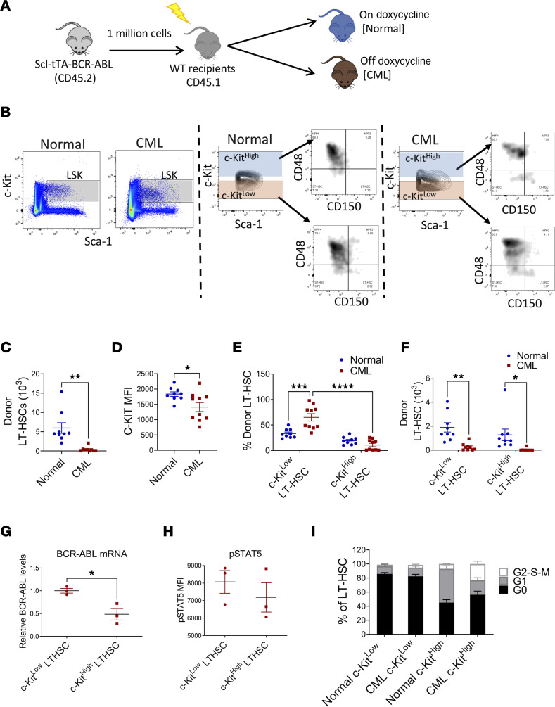
Figure 1. C-KITlo LT-HSCs are increased in CML compared with normal BM.
Experimental design: BM cells from SCL-tTA mice (CD45.2) were transplanted into lethally irradiated recipients (CD45.1) and were kept on doxycycline (normal controls), or without doxycycline, resulting in BCR-ABL expression and development of CML (CML). Mice were analyzed 10 weeks posttransplant (A). Representative FACS plots showing gating strategy for c-KITlo and c-KIThi LT-HSCs in normal (left) and CML (right) mice (B). Total number of donor LT-HSCs in the BM (C). Surface c-KIT expression (MFI) on donor LT-HSC in normal and CML mice (D). Frequency (E) and absolute number (F) of donor c-KITlo and c-KIThi LT-HSCs in normal and CML mice (n = 9–10). qRT-PCR analysis of Bcr-Abl1 mRNA expression in donor c-KITlo and c-KIThi LT-HSCs in CML mice (G). Results are expressed relative to GAPDH and normalized to c-KITlo LT-HSCs. Levels of p-STAT5 expression in c-KITlo and c-KIThi LT-HSCs from CML mice measured by flow cytometry after intracellular labeling with anti–p-STAT5 antibody (H). Cell cycle analysis of freshly isolated BM cells showing percentage of G0, G1, and S-G2-M phase normal and CML c-KITlo and c-KIThi LT-HSCs (n = 4 each) (I). Compiled data are presented as mean ± SEM, *P < 0.05, **P < 0.01, ***P < 0.001, ****P < 0.0001, based on t test (C, D, and H) and 2-way ANOVA with Tukey’s test (E and F).

