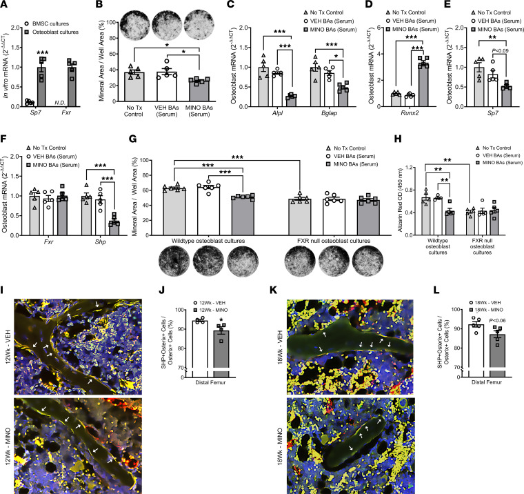
Figure 10. Minocycline-induced alterations in serum bile acids suppress osteogenesis through attenuating osteoblast/FXR signaling.
(A–E) Bone marrow stromal cells (BMSCs) were isolated from untreated 10-week-old female C57BL/6T specific pathogen–free (SPF) mice. (A) BMSCs were cultured in base media (α-MEM, 10% FBS, 1% PSG) versus osteogenic media (base media, 50 mg/mL ascorbic acid, 10 mM β-glycerophosphate) to evaluate differences in pre-osteoblastic cells versus mature osteoblastic cells. qRT-PCR: Sp7 and Fxr; n = 5/group. Unpaired 2-tailed t test; reported as mean ± SEM; ***P < 0.001. (B–F) Mature osteoblasts were stimulated with no treatment control (No Tx Control) or the altered serum bile acid profiles from minocycline-treated (MINO BAs) versus vehicle-treated (VEH BAs) female SPF mice; n = 5/group. (B) von Kossa assay; representative images and mineral area per well area (%) analysis. qRT-PCR: (C) Akp2/Alpl, Bglap/Ocn, (D) Runx2, (E) Sp7, (F) Fxr, Shp. One-way ANOVA with Tukey’s post hoc test; reported as mean ± SEM; *P < 0.05, **P < 0.01, ***P < 0.001. (G and H) BMSCs were isolated from untreated 10-week-old female C57BL/6J FXR-knockout and wild-type mice. Mature osteoblasts were stimulated with no Tx control, MINO serum BAs, or VEH serum BAs; n = 5/group. (G) von Kossa assay; representative images and mineral area per well area (%) analysis. (H) Alizarin red assay, optical density (OD) 450 nm. Two-way ANOVA with Tukey’s post hoc test; reported as mean ± SEM; **P < 0.01, ***P < 0.001. (I–L) Female C57BL/6T SPF mice were administered vehicle control (VEH) or minocycline (MINO) from age 6 to 12 weeks; euthanized at (I and J) age 12 weeks and (K and L) age 18 weeks. Immunofluorescence analysis of dual-labeled SHP+osterix+ cuboidal osteoblasts lining trabecular bone in the distal femur (green, SHP-FITC; red, osterix–rhodamine; blue, DAPI); n = 4/group: (I and K) representative images (original magnification, 200×), arrows indicate SHP+osterix+ osteoblasts; (J and L) SHP+osterix+ cells per osterix+ cells (%). Unpaired 2-tailed t test; reported as mean ± SEM; *P < 0.05 vs. VEH.

