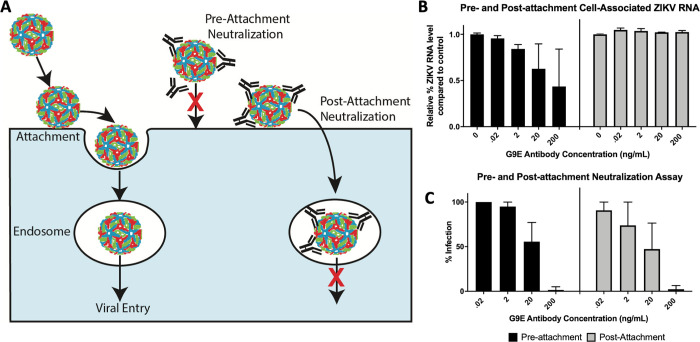
Fig 4. MAb G9E blocks steps in entry after ZIKV attachment to cells.
(A) Schematic of ZIKV entry into the target cell. After attachment to the cell surface, the virus moves to endosomes, where a drop in pH triggers membrane fusion and viral RNA release to the cytoplasm. Neutralizing mAbs can block steps before (pre-attachment neutralization) or after (post-attachment neutralization) virus attachment to the cell surface. (B and C) The mechanism of G9E neutralization was assessed by adding the mAb before or after the virus attached to cells. In the pre-attachment neutralization assay, ZIKV and different quantities of G9E were incubated together before adding the virus to cells (black). In the post-attachment assay, ZIKV was allowed to attach to the cell surface before adding different quantities of mAb (grey). Cells were harvested to measure the initial amount of virus that bound to the cell surface in the presence of the antibody (B) and the number of cells that were productively infected 24hrs later (C). Two independent experiments were performed to determine levels of cell surface associated virus (B) and four independent experiments (C) were performed to determine the percentage of infected cells. Results from representative single experiments (mean of technical duplicates) are depicted in panels B and C. Error bars represent the standard deviation of the mean (SD).

