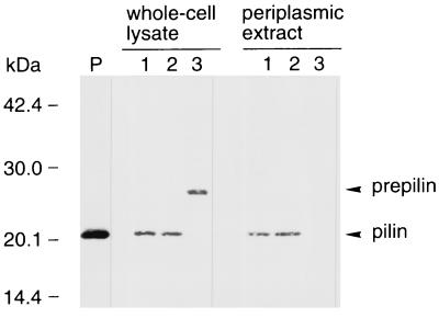
FIG. 2.
Western blot analysis of E. coli HB101 whole-cell lysates (left) and periplasmic extracts (right) using anti-CFA/III antiserum. Lane P, purified CFA/III; lane 1, E. coli HB101 harboring pTT202 and pTT224; lane 2, E. coli HB101 harboring pTT201 and pTT206; lane 3, E. coli HB101 harboring pTT202. The prepilin and processed pilin bands are indicated by arrowheads. Molecular mass markers are noted on the left side.

