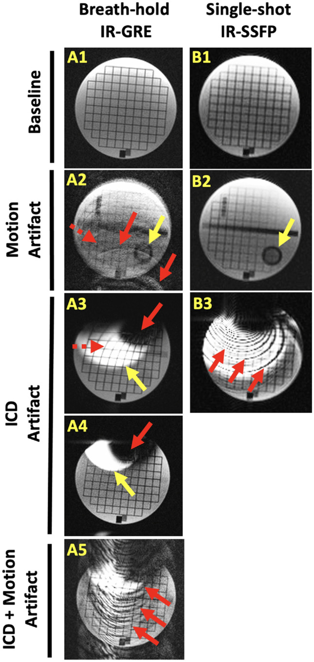
Fig. 1.

Illustration of motion and ICD artifacts for conventional LGE MRI techniques used for scar imaging. A1–5 Cardiac gated, breath-hold inversion-recovery (IR) gradient recalled echo (GRE) imaging collected with simulated heart rate 75 bpm. A1 image without motion is sharper than A2 with 2 cm, 3 Hz motion simulating poor breath-holding. Motion causes ghosting artifacts (e.g., red arrows), blurring (e.g., dashed red arrow), and appearance of structures that fall outside the desired image plane (e.g., yellow arrow). A3 placement of an ICD over the stationary object introduces different image artifacts caused by ferromagnetic ICD components. Hyperintensity occurs in areas that fall outside the bandwidth of the IR-pulse (yellow arrow). Distortion of the grid (dashed red arrow) and signal void (red arrow) are seen closer to the ICD, A4 Wider-bandwidth IR reduces hyperintensity artifact (yellow arrow). Higher receiver bandwidth additionally decreases image distortion closer to the ICD. A5 In the presence of an ICD, conspicuous ghosting artifacts are seen during 2 cm simulated respiratory motion (red arrows). B1–3 Single-shot IR balanced steady state-free precession (SSFP) imaging. B1 image without motion has lower image resolution and increased image noise compared to breath-hold imaging. B2 However, with 2 cm simulated respiratory motion, images have less blurring and ghosting compared to the breath-hold imaging. Still, objects outside the desired image plane can be seen (yellow arrow). B3) SSFP imaging is less useful for imaging subjects with ICDs due to significant banding artifacts
