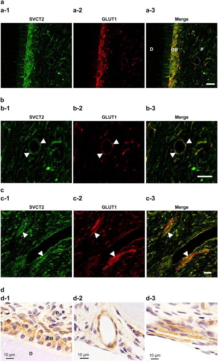
Figure 2.
SVCT2 was colocalized in glucose transporter 1 (GLUT1) in odontoblasts, blood vessels, and nerve fibers in normal pulp tissue. Double immunofluorescence staining of (a-1, b-1, and c-1) SVCT2 (green) and (a-2, b-2, and c-2) GLUT1 (ted). (a-3, b-3, and c-3) Merged images. (b-1–b-3) The arrowheads indicate the blood vessel. (c-1–c-3) The arrowheads indicate the nerve fibers. (d) Immunochemical stating of GLUT1. D, dentin, OB, odontoblast, P, dental pulp. Scale = 20 μm.

