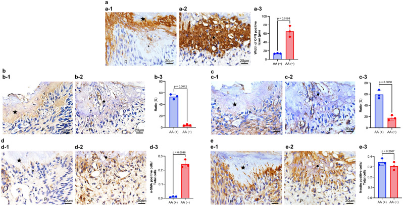
Figure 5.
Immunohistochemical alteration of osteopontin (OPN), type I collagen (Col I), type III collagen (Col III), α-smooth muscle actin (α-SMA), and Nestin in the pulp tissue of ODS rat 7 days after pulpotomy followed by MTA capping. Immunohistochemical staining of (a) OPN, (b) Col I, (c) Col III, (d) α-SMA, and (e) Nestin. (a-1, b-1, c-1, d-1, and e-1) ODS rats fed a normal diet (control; group 1). (a-2, b-2, c-2, d-2, and e-2) ODS rats fed an ascorbic acid-free diet (AA deficiency: group 2). Data exhibit quantification of OPN, α-SMA, and Nestin (a-3, d-3, and e-3), or rations of Col I and Col III (b-3 and c-3). Bars exhibit mean values ± standard error of the mean compared with controls (group 1), (n = 3 for each group; only comparisons with p-value ≤ 0.05 are shown), Welch’s t-test. The OPN-immunopositive layer is observed above the reparative dentin in group 1 (a-1), whereas the thick OPN-positive (necrotic) layer is found in group 2 (a-2). The OPN layer of group 2 is thickened in the necrotic layer (a-3). The reparative dentin is positive for the Col I immunoreactivity of group 1 (b-1), although Col I immunoreactivity is diminished in the necrotic layer and the pulp tissue of group 2 (b-2). The Col I occupancy of group 2 is diminished in the reparative dentin (b-3). The pulp tissue beneath the pulpotomy site is positive for Col III of group 1 (c-1), although Col III immunoreactivity is diminished in the pulp tissue (c-2). The Col III occupancy of group 2 is diminished beneath the reparative dentin when compared with that in group 1 (c-3). Alpha-SMA immunoreaction is faint or almost none under the reparative dentin (d-1), although α-SMA-immunopositive cells of group 2 are observed beneath the necrotic layer (d-2). The α-SMA-immunopositive cells of group 2 are increased in odontoblast-like cells when compared with those in group 1 (d-3). Nestin-immunopositive cells are observed along the reparative dentin in group 1(e-1) or under the necrotic layer in group 2 (e-2). The Nestin-immunopositive cells are unchanged in odontoblast-like cells between groups 1 and 2 (d-3). The closed star indicates the reparative dentin layer. The closed circle indicates the necrotic layer.

