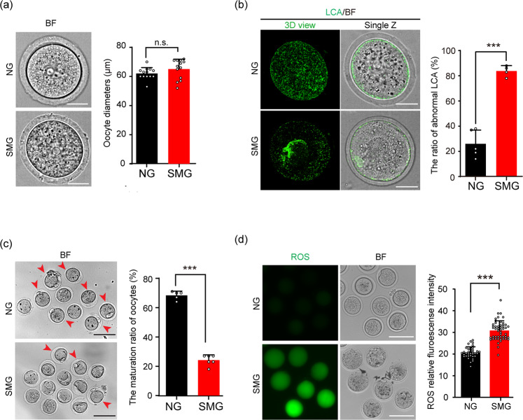
Fig. 2. SMG treatment decreased the quality of cultured follicle released oocytes.
a After follicles developed under SMG or NG conditions, the oocytes were isolated from the cultured follicles. No significant changes in the diameters of oocytes were seen when comparing the SMG group (n = 13) with the NG group (n = 13) after 2 days of follicle culture. p value = 0.18. Scale bars, 30 μm. b LCA (Lens Culinaris Agglutinin)-FITC immunostaining showing abnormal cortical granule distribution in the SMG group oocytes (n = 53) compared to that in the NG group (n = 59). p value = 0.000067. Scale bars, 30 μm. c Oocytes obtained from antral follicles after 16 hours of culture with LH in vitro, showing a significantly decreased ratio of PB1 (red arrowheads) in the SMG group (n = 74) compared to that in the NG group (n = 63). p value = 0.0000000031. Scale bars, 100 μm. d An increased fluorescence intensity which represented higher ROS level in oocytes of the SMG group (n = 50) compared to that in the NG group (n = 36). p value = 0.00000000000000000000078. Scale bars, 100 μm. Representative images are shown. Data are presented as the mean ± SD. Data were analyzed by two-tailed unpaired Student’s t-test and n.s. P ≥ 0.05, **P < 0.01, ***P < 0.001.

