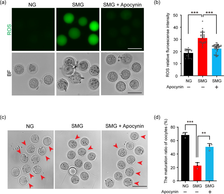
Fig. 6. Apocynin rescued SMG-related oocyte damage by decreasing the ROS level.
a After follicle culturing with Apocynin under SMG condition, the follicle-released oocytes showed a dramatically decreased ROS level compared to the oocytes without Apocynin. Scale bars, 100 μm. b The statistical analysis of DCF fluorescence intensity, showing a decreased ROS level in oocytes of the Apocynin group (n = 22 in NG, n = 26 in SMG and n = 41 in SMG + Apocynin). NG v.s. SMG: p value = 0.00000000000010, SMG v.s. SMG + Apocynin: p value = 0.000000028. c The ratio of PB1 (arrowheads) was markedly increased in the Apocynin-treated group compared to that in the SMG group. Scale bars, 100 μm. d Statistic analysis showing a significantly increased ratio of PB1 in the SMG + Apocynin group compared to that in the SMG group (n = 38 in NG, n = 35 in SMG and n = 43 in SMG + Apocynin). NG vs SMG: p value = 0.00021, SMG vs SMG + Apocynin: p value = 0.0019. Representative images of oocytes are shown. Data are presented as the mean ± SD. Data were analyzed by two-way ANOVA and n.s. P ≥ 0.05, **P < 0.01, ***P < 0.001.

