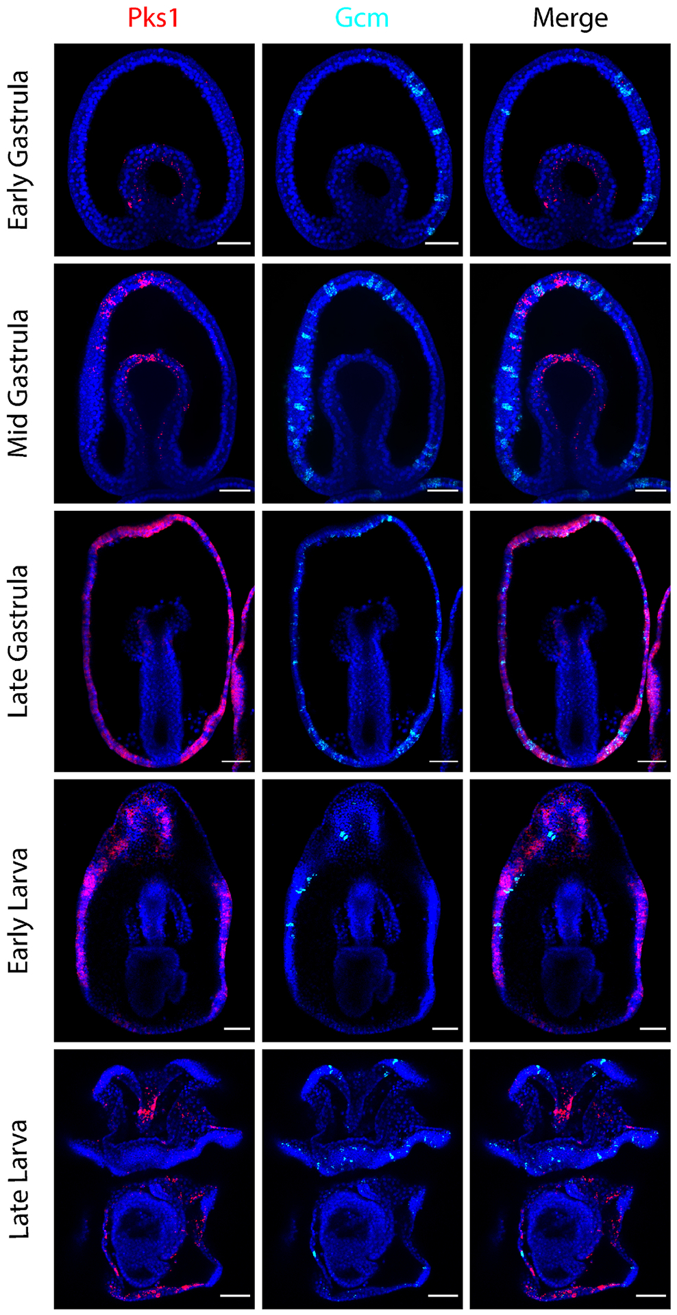
Figure 3. PmPks1 and PmGcm are expressed in distinct cells throughout development.

Confocal images show the expression pattern of PmPks1 and PmGcm using double FISH. As previously reported [47],[48], PmGcm is expressed by cells in the ectoderm throughout development. By the late larval stage, PmGcm-expressing cells appear mainly in the ciliary bands. At early stages of development, PmGcm is expressed in the ectoderm while PmPks1 is expressed in the mesoderm. Following the late gastrula stage transition in which PmPks1 expression appears in the ectoderm, PmPks1 and PmGcm still do not coexpress. Nuclei are shown in blue with DAPI. Scale bars are 50 μm.
