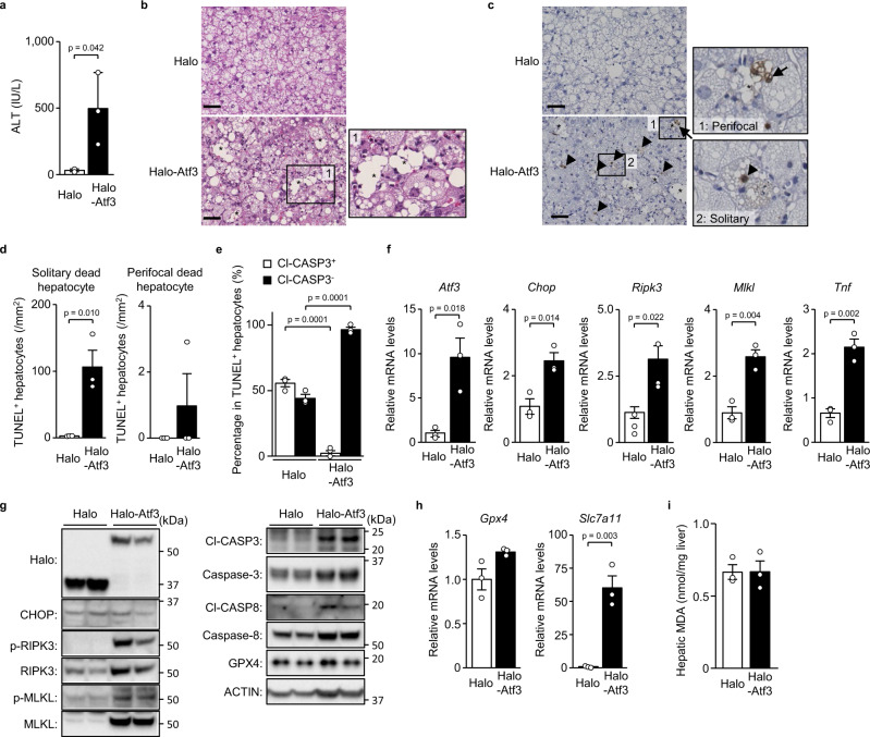
Fig. 4. Atf3 overexpression increases necroptosis in un-hepatectomised severe steatosis.
Halo-Atf3 or halo was overexpressed by adenovirus in mice fed a HFD for 16 weeks. a Plasma ALT levels. b Haematoxylin-eosin staining. Scale bar, 50 μm. Asterisks indicate hepatocellular death foci. c TUNEL staining. Scale bar, 50 μm. Arrowheads indicate solitary dead hepatocytes. Asterisks indicate hepatocellular death foci. Arrows indicate perifocal dead hepatocytes. d Number of TUNEL+ hepatocytes defined as described. e Cl-CASP3+ or Cl-CASP3− hepatocytes as a percentage of TUNEL+ hepatocytes determined by TUNEL/Cl-CASP3 double staining. f Quantitative PCR analysis of genes related to the eIF2α signalling pathway and necroptosis. g Immunoblot analysis. h Quantitative PCR analysis of genes related to ferroptosis. i Hepatic MDA levels. Data are presented as the mean values ± SEM. [n = 3/group, biologically independent samples]. Statistics: two-tailed Student’s t test (a, d–f, h, i). Source data are provided as a Source Data file.

